Imaging the replication of single viruses: lessons learned from HIV and future challenges to overcome
Susana Rocha, Jelle Hendrix, Doortje Borrenberghs, Zeger Debyser, Johan Hofkens (see publication in Journal or in Research Gate )The submitted version of this article can be obtained here.
Abstract
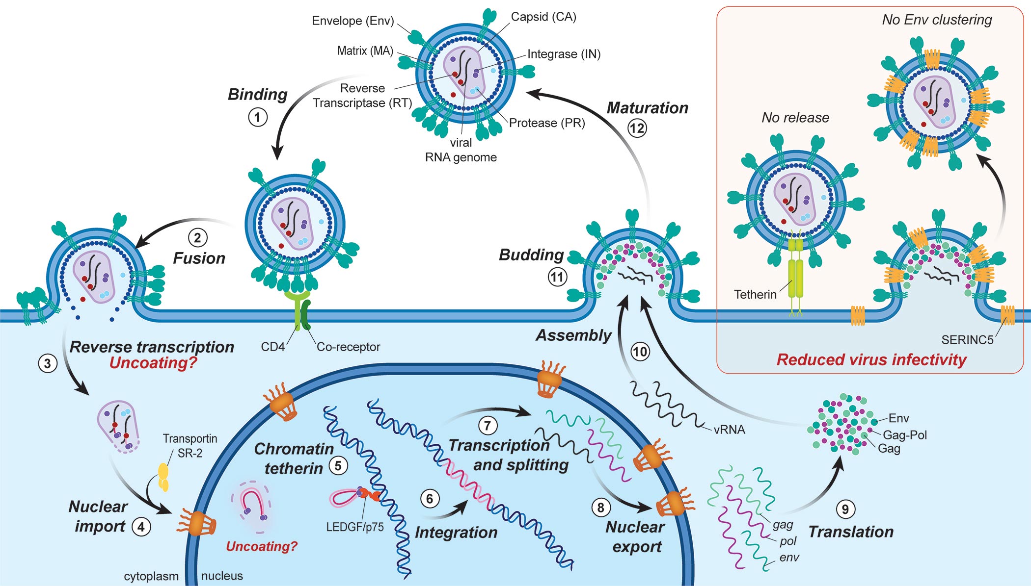
The molecular composition of viral particles indicates that a single virion is capable of initiating an infection. However, the majority of viruses that comes into contact with cells fails to infect them. To understand what makes one viral particle more successful than others, one needs to visualize the infection process directly in living cells, one virion at a time. In this perspective, we explain how single virus imaging using fluorescence microscopy can provide answers to unsolved questions in virology. We discuss fluorescent labeling of virus particles, resolution at the sub viral and molecular level, tracking in living cells, and imaging of interactions between viral and host proteins. We end this perspective with a set of remaining questions in understanding the life cycle of retrovirus, and how imaging a single virus can help researchers addressing these questions. While we use examples from the HIV field, these methods are of value for the study of other viruses as well.