Page Not Found
Page not found. Your pixels are in another canvas.

A list of all the posts and pages found on the site. For you robots out there is an XML version available for digesting as well.
Page not found. Your pixels are in another canvas.
Published:
Download here
Published:
Download here
Over the past decades, impressive breakthroughs have been made in cancer therapies. Namely, the development of multifunctional nanomaterials with enhanced efficacy and reduced side effects has marked a significant step forward in cancer research. However, the promising results observed in preclinical studies show low translatability into the clinic, due to a lack of adequate preclinical models that faithfully mimic the complex tumor microenvironment. Cancer research and drug screening are traditionally performed on 2D in vitro monolayer cultures and subsequently validated in in vivo animal models. Nonetheless, oversimplistic 2D cultures do not accurately represent the physiological 3D tumor architecture and lack adequate cell–cell and cell–matrix interactions. Meanwhile, animal models fail to properly replicate the human tumor microenvironment and are associated with high variability, costs, and ethical limitations. The 3D cell culture systems are a growing area of research to bridge the gap between traditional 2D drug-screening platforms/animal models, and the human body. These tridimensional cancer models better recapitulate in vivo tumor features and have demonstrated more clinically relevant therapeutic responses in comparison with 2D and animal models. This chapter provides a comprehensive overview of the available 3D cancer cell models to date, including scaffold-free and scaffold-based techniques for tumor spheroid formation, a description of patient-derived tumor organoids/tumoroids, and 3D microfluidic-based strategies. In doing so, we pinpoint each model's main advantages and disadvantages and highlight their relevance as preclinical models for nanomedicine.
Published:
Download here
Published:
Cancer is one of the major causes of death in our society. The anti-cancer drugs currently used in chemotherapy produce several side effects and, although fatal for almost all cells in a tumor, a small percentage of cells appear to resist the treatment. It is therefore urgent to design new therapies that specifically target these cells while causing no harm to healthy tissue. The resistance to multiple drugs is linked to the presence of specific molecules at the cellular plasma membrane, that actively pump out chemical drugs (efflux pumps). Additionally, some cancer cells have the ability to self-renew, thereby initiating secondary tumor growth. Multi-drug resistant cells and cancer stem cells are suggested to be the main cause of treatment failure and relapse.
Published:
Published:
bla bla bla
Published:
A website to share the cool science we do at KU Leuven.
Published:
Congragulations Wout & Willem! Nice article reporting new development in correlative AFM and Fluorescence imaging, and its application to study the interaction between DNA and DNA-interacting proteins. The full article can be found here.
Published:
Congragulations Max! Finally some good news from the EMBO organization: Max received the EMBO travel grant for the 3-month stay in Leuven. He will be working on imaging focal adhesions in 3D (more information on this project can be found here)
Published:
Congragulations Lieve & Koen!
After some time, this story is (finally) out. A nice collaboration between virology, microscopy and photophysics experts, reporting a phenomena that will give a lot to talk about… The full article can be found here.
Published:
Congragulations Nagma, Doortje & Jelle!
We were able to write a nice article reviewing our work in the virus field. The full article can be found here.
Published:
Congragulations Indra & Boris! Awarded FWO doctoral grant. It is going to be 4 years of fun with Nanoparticles & Polymers!
(Unfortunately, I do not have such a pretty foto of Boris…)
Published:
Congragulations Guy! More than 2 years after submission, this story is finally out! I am proud of my small contribution. The full article can be found here.
Published:
Congragulations Herlinde! I am very proud of this one. After all the struggle with the cloning, and all the re-writing to make the best story possible, 2 years after the PhD - it is out! With the final contribution of Dr. Rafael Camacho and his nice simulations. The full article can be found here.
The software to run the simulations can be found here.
Published:
Colaboration Leuven - Madrid keeps increasing!
Thanks to EMBO I will be able to join Guillermo Solis and Dr. Rodrigo Barderas at the Instituto Salud D.Carlos III in Madrid. I am really looking forward to meet the Barderas’ group and get this project going!
Published:
After an unforgettable Xmas party, a few days to (try to) finish up things and we are off to a well deserved Xmas break! 2019 might bring big changes - let’s hope they are all for the better!
And if not, let’s just continue the spirit and do great science!
Happy new year!
Published:
Even though we were not directly involved, the Leuven team is very happy to see this work published. Congratulations Max! All your hard work is starting to pay off. This is the first one, hopefully many more will follow.
The full publication can be found in the journal or at Research gate.
Published:
Congratulations Hongbo!
Dr. Hongbo Yuan will be joining us after the summer, to investigate forces from the cell or the ECM change the mechanical properties of the matrix and, more importantly, how this change affects biological functions. We are really looking forward to work with you on this.
Published:
A nice article showing how simple chemistry can improve cancer specificity and efficiency of drug delivery systems.
Congratulations Bea!
You were also able to (finally) show that PEI does induce endossomal escape. And, for the non-believers, more evidence will follow!
The full publication can be found in the journal or at Research gate.
Published:
We could have ordered pens or mugs but… the amount of sun in Belgium requires protection!
An item to give to current and future lab members.
(now we have to wait for the end of the summer to join the team for a group picture)
Published:
Thanks to Dr. Nuno Canha, I could present my research to colleagues from my home country, including some good friends.
It was a short visit, but a memorable one.
The quality of the science being done in countries with very limited resources will always amaze me. For good science, you need a good brain, with a substantial amount of creativity.
Published:
Today was a day of many fruitful discussions with visitors from Hokkaido University.
I had the opportunity to give a talk presenting our research, while Johannes and Quinten could present their progress during the poster session.
We have found some common interests in fluorescence-based sensors, drug nano delivery systems and extracellular matrix. Next meeting in Japan - already looking forward!
Published:
Today we celebrated the 80th anniversary of Prof. Frans De Schryver.
The list of speakers included Ben Feringa, Tanja Weil, Roeland Nolte, Klaus Müllen, Jürgen Rabe, Hiroshi Masuhara, Thomas Ebbesen, Loredana Latterini, Paolo Samori, Markus Zauer and Hua Zhang.
A day full of interesting talks, with the additional pleasure of reviewing old colleagues.
Published:
Today we celebrated the 80th anniversary of Prof. Frans De Schryver.
During the symposium in honour of Prof. Frans De Schryver, there were 8 poster awards.
Congratulations Johannes, Monica, Beatrice and Marisa! 👏 👏 👏 👏 👏
Published:
The Marie-Curie project of Dr. Hongbo Yuan starts with a visit to his former lab, Kouwer’s group at the Radboud University in Nijmegen.
A collaboration that started 2 years ago, with the visit of Prof. Rocha. After that, Khaizheng Liu (Max) visited our lab, Johannes Vandaele visited Nijmegen, Dr. Hongbo Yuan got a competitive Marie-Curie fellowship and soon Dr. Röel Hammink will come for a short stay.
A good example how collaboration between group with different background can drive science forward.
Published:
We decided to celebrate the start of Rocha’s lab with a nice team building day. After a tiring and exciting GPS game of cops and thieves (some better than others), we made peace during dinner. Congratulations to Boris, Laurens and Elizabete for their win (but next time we will catch you!)
Published:
After some attempts, Johannes was able to convince the FWO expert panel of his project (and his personal abilities as a scientist). Congratulations Mr. FWO_aspirant!
A good example where hard work and persistence paid off. (and now still 4 more years ahead!)
Published:
EMBO will support the visit of Dr. Hammink to our laboratory. While he is also working with synthetic polymers, he uses this system to activate T-cells. Very interesting work from the group of Prof. Carl Figdor (Radboud University, Nijmegen).
We will use our cutting-edge microscopy techniques to characterise the interaction between polymers and immune cells. I am confident that his visit will result in top science.
Published:
It is strange to return to your university 15 years later to give a seminar and see some of your old professors in the room. Thank you Prof. Gaspar Martinho for the invitation. A day or very useful discussions and hopefully the first meeting of a long standing collaboration on bio-applications of nanoparticles. The work done by Prof. Carlos Baleizão and Prof. Paulo Farinha on tailored design of nanoparticles is amazing! Looking forward to get our hand in those particles!
Published:
Congratulations Ovia! An very nice article reporting the best FRET acceptor for NanoLuc.
Let’s hope this will be the first of many successful research projects.
The full publication can be found in the journal or at Research gate.
Published:
Congratulations Hongbo! A nice example of a biological application of an hybrid hydrogel.
The full publication can be found in the journal or at Research gate.
Published:
The PhD is hosted by the Rocha lab at KU Leuven, which develops fluorescence microscopy tools and assays to address biologically relevant questions. The group works at the crossroad between biology, chemistry and nanotechnology. The main imaging modalities use are confocal, multi-photon, single molecule fluorescence (single particle tracking, super- resolution) and multi-plane wide field microscopy. In parallel, we work on the optimization of biomimetic matrices for 3D cell models and functionalization of nanoparticles.
Published:
For years we have talk about doing a lab trip to Lisbon. This year, we the excuse of celebrating the start of the ‘Rocha Lab’, we finally did it! A lot of walking, eating, drinking, and above all, laughing. It is an amazing group of people. Ah yes, Lisbon is an amazing city also.
Published:
Congratulations Marisa! All those hours/days/weeks bleaching squares finally paid off.
The full publication can be found in the journal or at Research gate.
Published:
Congratulations Johannes!
Published just before the COVID-19 lockdown (apologies for the delay in the congratulations). It is the first article of Joahnnes and the first of our collaboration with the group of Paul Kouwer. Let’s hope it is the first of many!
The full publication can be found in the journal or at Research gate.
Published:
Pierre is joining us as one of the two PhD students that will be working for the SuperCol project at KU Leuven. A highly interdisciplinary project, requiring very specific skills. Pierre, with his unique background, has impressed us since the first interview. Looking forward to get started!
Bienvenue Pierre!
(There is no ‘NanoGirls’ anymore, we are now a ‘NanoTeam’!)
Published:
Congragulations Bea and Farsai! The article where we used FRET to investigate the intracellular degradation of nanoprodrugs has been published in Nanoscale. Check the publication (here)
Published:
Single molecule imaging has been crucial to unravel molecular mechanisms of several biological systems. In this perspective, we tried to summarise how imaging single virus particles has contributed to the understanding of the HIV viral cycle. It a pleasure to write this article with the Prof. Zeger Debyser, Prof. Johan Hofkens, Prof. Jelle Hendrix and Dr. Doortje Borrenberghs.
Published:
It was a long road from ‘Let’s just publish how we do this’ to the final article. But it was worth it!
Congratulations Boris! Now that the method is published, it is time to move on and do some ‘real’ science!
(But first, some drinks!)
Published:
We started writing the review before COVID arrived in Belgium, but it will always be remember like the review we wrote during the lockdown. The goal was to summarised all the information about using nanoparticles in 3D cell culture systems, and create a small guide for researchers with limited knowledge of biology.
I am really proud of this article! #girlPOWER
Published:
We were awarded with a project within the “KU Leuven Global PhD Partnerships with Melbourne University” program. The project, entitled “Advanced cell models and multifunctional nanomaterials for light- mediated cancer therapies“ is a collaboration with the group of Prof. Paul Mulvaney and Dr. James Hutchison (a previous post-doctoral researcher of KU Leuven, an a friend).
The PhD position in Melbourne is already open, the position in KU Leuven will open in April/May.
More info? See the project description
Published:
Congratulations Monica! It was not always an easy path, but you have reach the end (or should I stay a crossing?).
The hours of measuring, the days of sample preparation, the challenges of interdisciplinary research... A PhD defence and a paper accepted on the same day.
We wish you all the luck with your future endeavours!
Published:
Sam has joined the group as a new PhD student. He will strengthen the FRET-based sensors team and has the ambitious plan of taking FRET-based force sensing to the third dimension, using 3D cell models and micro-fluidic platforms. More details on his project can be read here.
Welcome to the team Sam!
Published:
Congratulations are in order for Dr. Laskaratou! After successfully publishing a nice article in Nature Communications (see the publication here.), Danai successfully defended her PhD thesis. Luckly, the current regulations allowed a hybrid defence, and Danai could present her research to the jury members, friends and family. Her explanation of fluorescence, FRET and cell signalling in layman’s terms was superb!
Danai also wrote a nice ‘behind the paper’ contribution, which you can read here.
Published:
We are organising a summer school to teach early stage researchers how to use fluorescence microscopy to investigate cellular forces. A mix of lectures, tutorials & hands-on workshops. Organised in collaboration with the group of Prof. Van Oosterwyck (KULeuven) and with the support of Arenberg Doctoral School and PicoQuant. Interested? Find out more here.
Published:
Congratulations Bea, Monica - and all the co-authors!
Endoscopy is a very challenging tehcnique but this publication demonstrates how powerfull it can be. Optimized nanowire-based endoscopy probes were used to monitor pH changes in living cells, in both the cytoplam and the nucleus.
The full publication can be found in the journal or at Research gate.
Published:
Very proud of organzing this AMAZING summer school! The first time - and immediately a huge sucess. Three days of lectures, tutorials and workshops. Special thanks to Prof. Paul Kouwer, Prof. Jelle Hendrix and Dr. Mar Cóndor for their contribution.
Published:
With the start of the academic year, I would like to welcome the new students that joined the group: Silke, Maria, Astha and Sarah!
(Also a late welcome to Ana, who arrived already in the beginning of the year)
RochaLab(2021) = RochaLab(2020) + girls5 #WomenInSTEM
Published:
Congratulations Marisa! You made it! After 4 years of hard work, you reach the the light at the end of the tunnel...
. Next challenge: expanding your knowledge in the medical sciences. I am 100% sure you will continue to make significant contributions to the advance science.
I hope to see you around!
Published:
Like this it is fun to get older!
A big ‘Thank you’ to the team for the presents, they were perfect!
P.S. Drinkable gifts did not survive long enough to be photographed…
Published:
Congratulations Dr. Aline Acke
! A PhD on ‘expanding the unexpandable’. A very nice presentation, wanted by all promotors and co-promotors… I wish you all the luck in your new adventures, both on your professional and personal live.
Published:
The visit of the group of Prof. Paul Kouwer was an amazing day, where we shared lots of science and ideas with . Thanks for visiting the chemistry department.
We are already looking forward to the next meeting!
Published:
Congratulations Bea and Hongbo! Today we celebrate! Both Bea and Hongbo will receive a pretigious FWO fellowship to continue their work on nanoparticles and biomaterials. Three years of funding!
#HappyPI
Published:
The first edition of the Nanocenter Image Contest was a success!</b> With more than 40 participants, it was difficult for everyone to chose thier favorite...
Congratulations to the winners and let's see what the second edition will bring...
(and a special thanks to Rik Nuyts for the support on the organization)
Published:
It took some time and hard work, but Guillermo was able to finalize this nice story!
And publish it on the British Journal of Cancer!!! Congratulations!
The full publication can be found in the journal or at Research gate.
Published:
In this article, Aline applied Expansion microscopy to investigate the co-localization between epigenetic readers and markers for hetero- and euchromatic.
Just out-of-the-press, to show that it is possible to publish your work even after finishing your PhD.
Congratulations Aline!
The full publication can be found in the journal or at Research gate.
Published:
After 5 years of being around in the lab (PhD and master thesis), Indra sucessfully defended her PhD. She was one of a kind and will be missed.
Congratulations and thank you once again for starting 3D cell culture in the Rocha Lab.
We wish you all the luck on your next adventure!
Published:
After countless trips from Leuven to Madrid, and a almost endless pile of paperwork, Guillermo has finally able to defend his PhD in September (or will it be in November?..)
The jury agree: your work would be worht of summa cum laude if KU Leuven woudl allow distinctions on the doctorate degree.
Anyway, congratulations (x2)! Thank you for being such a giving person, helping everybody, and setting the work ethics really high in the lab.
Published:
In cold Sweden, Prof. Theo Lasser has placed Boris in icy grounds …. But he resisted, kept his coolness and triumphed! Another dual PhD degree, just to show how much our students like paper work!
Congratulations for the heroic effort and resilience! It was a truly unique defence, followed by an amizing party. Both reflected your character very well: a brillant and warm person!
Published:
Congratulations Johannes! For the PhD and for the amazing presentation you gave in Dutch!
We were left with mix feelings of being proud and happy for you, and a bit sadness for letting you go 'across the fence', to the industry. I am 100% sure you will succeed in whatever you set your mind into. You were without a doubt one of the most hard working students, even if the work was not always related to science ;)
Published:
Congratulations Hongbo, and all the other authors! This was truly a team effort, only possible in collaboration with Paul Kouwer’s group, and with the support of the Matrix group.
Several years later, we were finally able to perform TFM in PIC gels - and learn a lot from it!
The full publication can be found in the journal or at Research gate.
Published:
Congratulations Boris and Guillermo! 👏 This amazing duo was able to secure two prestigious FWO grants. Super happy to be able to count on them to explore optical trapping and cancer metastasis - and ensure that parties will keep going 😉
Published:
The second edition of ‘Feel the Force’ summer school is a wrap! 🔬 Big shoutout to all the participants for their interest and ‘mechano-passion’. 🙌❣️ Huge thanks to the Rocha Lab & Co for their incredible support! 😊 Couldn’t be happier with this awesome team! 👏
A special thanks also to the amazing speakers who accepted to participate! Prof. Jelle Hendrix, Prof. Xevi Casadevall i Solvas, Dr. Eva Faurobert, Prof. Aurélie Carter, Prof. Bart Smeets, Prof. Paul Kouwer and Dr. Abdel Fattah.
Published:
After 4 years of meetings, collaborations, scientific exchanges and a lot of fun, the SuperCol network reached its end… 😢 It was really a pleasure to work with this group of young scientists, and see them growing and become independent researchers.
I wish you all the best with your future careers!
Published:
Delighted to have hosted Hokkaido University’s scientific delegation at KULeuven for the Core-2-Core symposium yesterday! 🥳After 4 years due to COVID, it was fantastic to meet in person. Big thanks to our colleagues from Melbourne University for joining us online! 🌏
Published:
Congratulations to Quinten for defending his PhD! 🎉🎓 There was fluorescence imaging, some curve fitting, strange equations but above all, a lot of fun! You leave a legacy behind: a new research line on FRET-based tension sensors and, more important, coffee mugs for the group 😉 Enjoy your return to the motherland of West Flanders and keep in touch!
Published:
❄️ While red weather alerts sweep the nation, we’re brightening up the Chemistry department with Rocha Lab’s creative decorations!☃️ #TeamSpirit #LabLife
Published:
Johannes included on his PhD thesis a chapter on ‘all you need to know about PIC’, so that the lab members joining after him could quickly gather the most important information. With additional support of Max (Khaizeng Liu) and Roel, we manage to publish this work. We believe the material community will greatly benefit from this review.
The full publication can be found in the journal.
Published:
Super proud to share the success of the IUPAC Global Women’s Breakfast held at KU Leuven! With the support of the Chemistry department, we hosted over 60 enthusiastic participants. A big shoutout to our speakers for their openness during the Q&A session👩🔬 #WomeninSTEM
Published:
Following the successful duo Maria and Samantha, we are super excited to be awarded a second Global PhD Partnership. We will develop methods to probe and control the mechanical properties of synthetic ECM, at the subcellular scale.
It is was very special to develop projects with the other side of the world…
Published:
We are thrilled to announce that Indra Van Zundert has been awarded a prestigious post-doctoral fellowship from FWO! 🎉 She will be joining our team in October, using 3D cell models to develop DNA origamis for gene therapy. 🧬 Can’t wait to have you with us at KU Leuven!
Published:
Light therapy + nanoparticles = a powerful cancer-fighting duo!
Do you want to know more? Read our latest review on current advancements!
A nice collaboration between KU Leuven and University of Melbourne, with Maria Bravo, Beatrice Fortuni and James Hutchison taking the lead!
The full publication can be found in the journal.
Published:
🎉 Huge congratulations to Charlotte Cresens for very successfully defending her PhD 🎓 I am incredibly proud of seeing Charlotte achieve this incredible milestone, though it’s bittersweet knowing she won’t be in our lab anymore. 🥹
But, as she embarks on her next journey as a super-resolution microscopy expert at the VIB imaging core in Leuven, I’m certain our paths will cross again. 🥳 Best of luck, Charlotte, in your future career — you will do amazing things!
Published:
While I was not able to attend, Rocha Lab was present at the MAF 2024 in Valência.Guillermo and Samantha were selected for oral talks, Sarah, Sandra and Maria presented their latest results, and Steven return to Leuven with a Best Poster prize award 👏👏👏
#ProudPI
Published:
Nina Anseeuw outdid herself with this lab-logo masterpiece for her birthday.
Starting 2025 on a sweet note with science and great vibes!
Here’s to more good times (and good science)! 🔬✨
Published:
How do cells translate molecular forces into mechanical actions? 🧬🔬💪
Our new research combines Traction Force Microscopy (TFM) with FRET-based tension sensors to uncover how focal adhesions manage vinculin tension and cellular traction. Our findings highlight the dynamic interplay between molecular-scale tension and large-scale cellular mechanics.
Check it out here.
Massive thanks to Samet Aytekin for driving this work, with key contributions from Laurens Kimps and Prof. Hans van Oosterwyck!
Published:
What if tweaking nanoparticles isn’t enough to improve nanomedicine? 🧐 In our latest work, we show that tumor biology – not just particle design – affects how nanoparticles behave. Same particles. Different tumors. Totally different outcomes. 🤯 Big shoutout to Maria Bravo, Guillermo Solís, Beatrice Fortuni and all co-authors for pushing this forward! 🔎 Curious? Check it out here.
Published:
A rare gathering in Leuven…
For the first time, we brought together both generations of PhD students from our Global PhD partnership between KU Leuven and University of Melbourne:
Published:
Taking part in Wetenschap aan Zee 2025 was a wonderful opportunity to bring our research out of the lab and into the public sphere. I had the pleasure — and the challenge — of presenting our work on nanotechnology and mini-tumors to a general audience, entirely in Dutch. Many thanks to the organizers, and in particular to Prof. Steven De Feyter, for the invitation. It was equally inspiring to share the stage with Prof. Maarten Roeffaers, whose talk on detecting microplastics with light offered a fascinating look at science in action. Events like this remind us of the value of science outreach — sparking curiosity, encouraging dialogue, and making complex ideas accessible to everyone.
Published:
Cells sense physical forces and the mechanical properties of the microenvironment via several distinct mechanisms and cellular components. The first step of cellular adhesion to the ECM occurs via transmembrane heterodimers of the integrin family. Once integrin molecules adhere to the ECM, they are activated and form clusters. As the number of bound molecules increases, some of the focal complexes evolve from small (0.5-1µm in diameter) transient ‘dot-like’ contacts to elongated structures (3-10µm) which couple with actin and associated proteins. The mechanical coupling between the ECM and the cell cytoskeleton is controlled by the dynamics of the focal adhesion complexes (assembly, disassembly and turnover).
Tissue stiffness plays a critical role in regulating cellular behavior, significantly influencing processes such as cell migration, differentiation, proliferation, and apoptosis. Cells sense and respond to the mechanical properties of their environment through a process known as mechanotransduction, wherein mechanical signals are converted into biochemical responses. This process is largely mediated by integrins, focal adhesions, and the cytoskeleton, enabling cells to “feel” the stiffness of their surroundings and adjust their functions accordingly. In a healthy tissue environment, cells exist within an optimal range of stiffness that supports normal function. However, when tissues become abnormally stiff due to pathological conditions like fibrosis or cancer, cellular behavior can change drastically, often promoting disease progression.
Mechanical forces play an undisputed elementary role in the interactions between cells and the surrounding extracellular matrix (ECM) [1]. Not only are these forces essential for the cells migratory behavior, they also influence proliferation (including tumor growth) and differentiation [2–4]. These forces are transferred across focal adhesions (FAs) which connect ECM and cell skeleton through patches of activated integrin proteins. Since the origin of exerted cellular forces lies in these FAs, they are the ideal starting point for characterization of mechanotransduction pathways. Consequently, studying how the properties of the ECM affect the cellular forces is key in understanding how cells connect to their environment and alter their behavior appropriately. While the scientific field of cellular mechanosensation has been studied for years, recent developments in imaging techniques and force sensor development enable us to dig deeper.
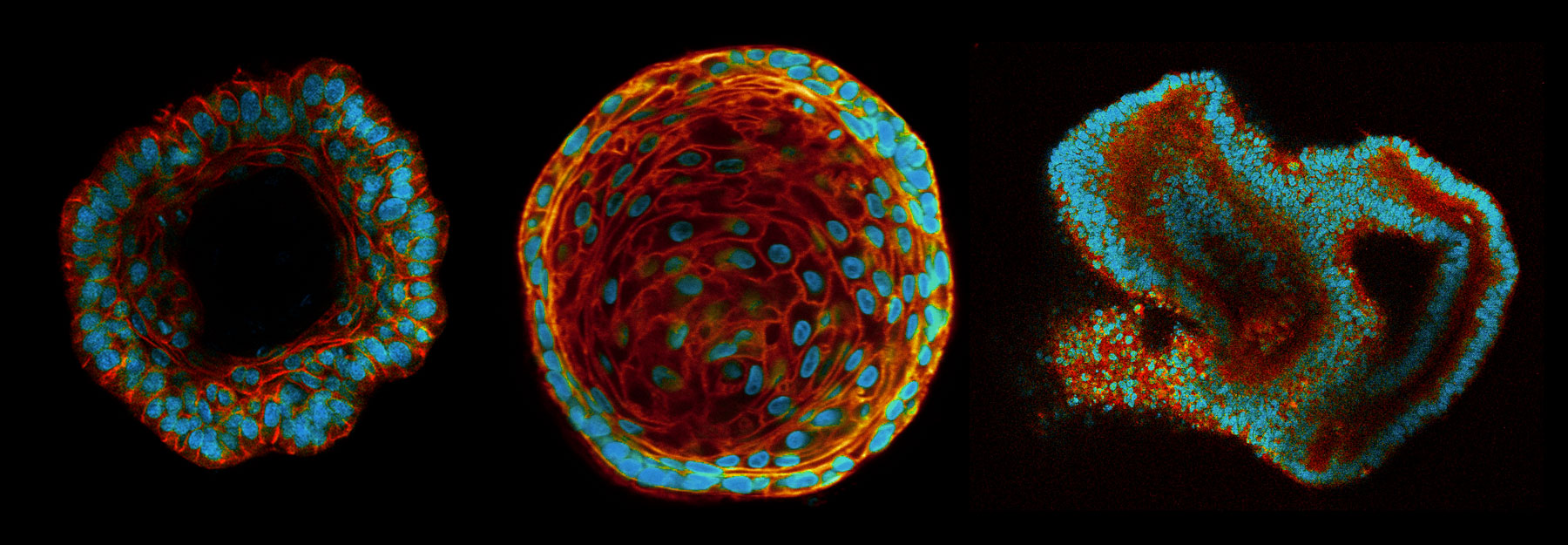
Nowadays, human organoids are becoming a highly promising tool to model organ development, function and especially human diseases in vitro. In general, organoids are miniature, simplified organs that can easily propagate in vitro originating from one or a few cells, typically stem cells.
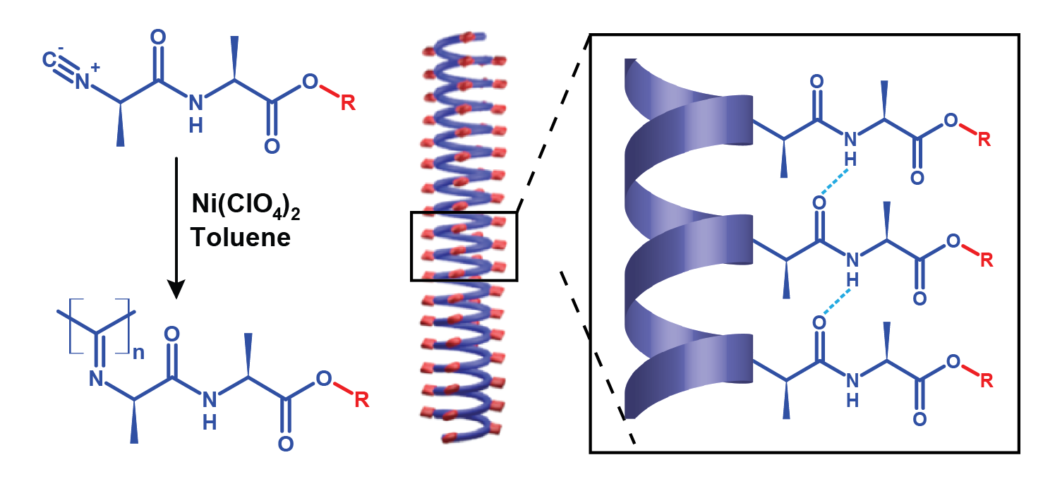
A large number of natural and synthetic hydrogels are currently used for tissue engineering and regenerative medicine. Over the last decade, there has been an increasing awareness of the role of material properties of the substrates in guiding cellular behaviour. This has inspired chemists to create a new generation of materials with mechanical properties closed to that of natural occurring biopolymer networks. Recently, the groups of Prof. Alan Rowan (Queens University, Australia) and Prof. Paul Kouwer (Radboud University of Nijmegen, The Netherlands) were able to develop a fully synthetic material that mimics in all aspects the gels prepared from cellular filaments. These synthetics gels are prepared from polyisocyanopeptides (PICs) grafted with oligo(ethylene glycol) chains and share structural features of biopolymers: their helical structure renders the polymer molecules relatively stiff while the interaction between the side chains enable the formation of bundles or fibrils of defined dimensions. The triethylene glycol side chains attached to the polymer backbone render the material thermo-responsive (it will gel upon heating beyond 20 °C and become liquid again upon cooling). Despite being characterized extensively in bulk, the fundamental dynamics and the relation between the macroscopic properties and the microscopic structure at cellular length scales of PIC-based hydrogels remains obscure.
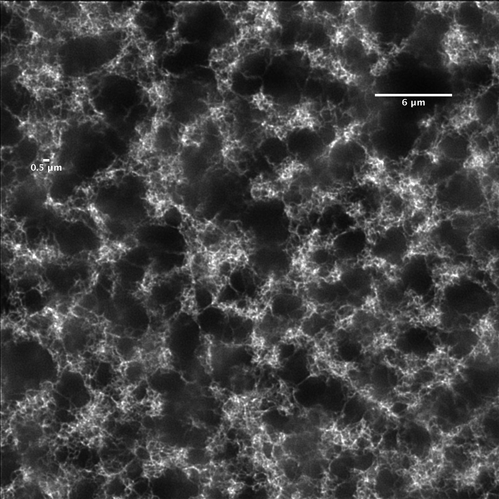
Classically, structural characterization of materials is performed with electron microscopy or scanning probe microscopy. Despite the high spatial resolution achievable with these techniques, they are unable to measure dynamics ‘in situ’ and sample preparation can be a laborious process. In contrast, optical microscopy has the potential to unravel the dynamics in complex heterogeneous systems but has been limited to a spatial resolution of ca. 200 nm. In the past 10 years fluorescence imaging has been revolutionized by the successful development of sub-diffraction (super-resolution) microscopy modalities which can achieve resolutions down to tens of nanometers (see Molecular Organization at the Nanoscale).The various possibilities of fluorescence microscopy to probe dynamics and heterogeneities, with molecular resolution, for a wide range of time scales makes it an ideal tool to address many topics of polymer science. In this project we are using fluorescence microscopy to image the polymer network at the micro to nanometer scale.
Our most recent results can be found in this publication.
For more information on PIC-based hydrogels:
Due to the crucial role of physical cues in regulating cell behaviour, the mechanical properties of hydrogels are a key design parameter in tissue engineering applications. The shear elastic properties of viscoelastic materials are commonly measured by mechanical rheometers. Storage and loss moduli of a material can be measured by application of strain while measuring stress or vice versa. In contrast, recently developed optical micro-rheology techniques use nanometer- or micrometer-sized particles embedded in the material to obtain the viscoelastic response parameters. Thermal or passive micro-rheology for viscoelastic materials is based on an extension of the concepts of Brownian motion of particles in simple liquids. The movement of the embedded particles can be monitored using particle tracking. Initially developed to investigate the rheological properties of uniform complex fluids, particle tracking micro-rheology (PTM) is becoming a popular technique to analyze polymer blends and gels, as well as the deformability and elasticity within cells. However, if the beads locally modify the structure of the gel or are contained in a pore in an inhomogeneous matrix, the bulk rheological properties will not be retrieved. A solution is to use the cross-correlated thermal fluctuation of pairs of tracer particles, ‘two-point micro-rheology’. This method provides a better agreement between micro and macro-rheology, even in complex micro-structured fluids. However, technical constrains limit the wide application of this technique. One of the major limitations of two-point micro-rheology is the reduced number of trajectories that can be used for analysis. During particle tracking micro-rheology, the length of the calculated trajectories is limited by the time spent by the tracers in the field of view (x,y) and depth of focus (z). Consequently, mechanical characterization of complex polymer matrixes at the micrometer scale would benefit greatly of a new method for (fast) tracking in 3D. We are developing a new method for fast tracking of (fluorescent) beads in 3D using a multi-plane wide field microscope. This will allow a better mechanical characterization of soft materials, at the microscale.
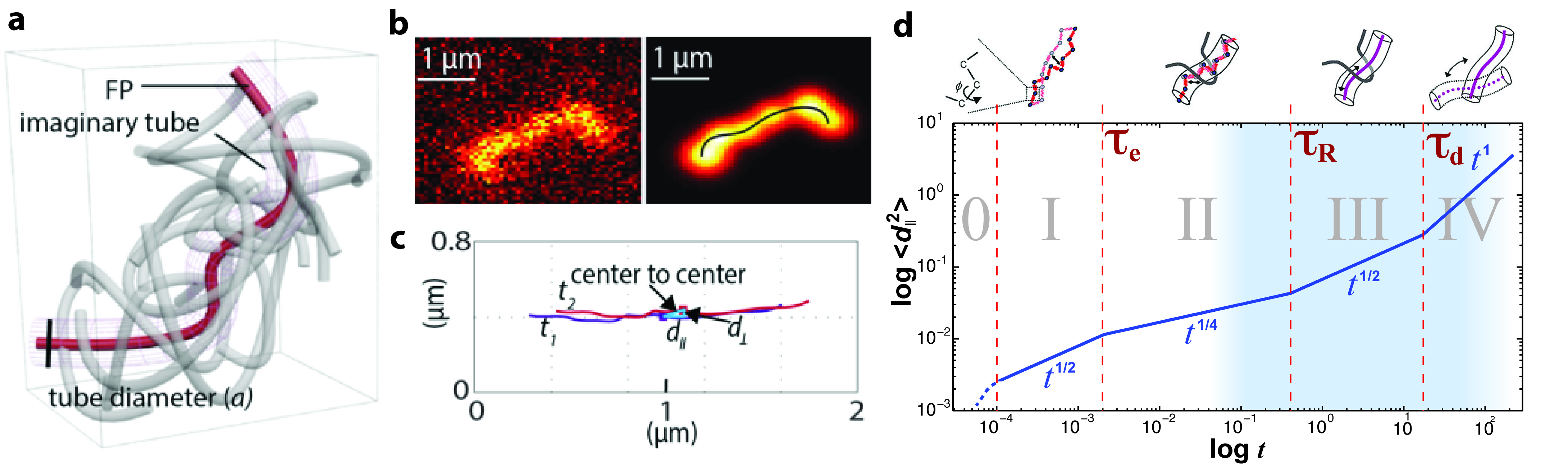
Our current theoretical understanding of entangled polymer chain dynamics is based on the reptation model. First proposed by Doi and Edwards, and further expanded by de Gennes, the reptation model assumes that a polymer chain is confined by the surrounding matrix and is therefore forced to move inside an imaginary tube defined by the transient network of entangled neighboring chains. Intuitively this motion resembles that of a snake or worm. The reptation model predicts five dynamical regimes for segment diffusion, summarized in the figure below. These regimes are as follows: (0) sub-segmental processes (“glassy dynamics”) at very short times (microseconds), (I) small motion subject only to chain connectivity, (II) “local reptation”: short-distance motion within the constraints imposed by the surrounding chains (“tube”), (III) “reptation”: diffusive motion along the curvilinear tube over distances larger than the polymer size, and (IV) free diffusion.
Since Ashkin’s breakthrough in 1986, optical trapping has been widely used to manipulate micro- and nanoscale objects across fields like material science and biology. The method typically involves using a tightly focused laser beam, where particles are attracted to the laser focus by the spatial gradient force.
Photothermal therapy (PTT) and photodynamic therapy (PDT) are innovative cancer treatments that harness the power of light to selectively target and destroy cancer cells, offering non-invasive alternatives to conventional therapies.
In this decade, the pharmacology field has been intensively exploring different approaches to deliver multiple drugs with a single drug nano-carrier, such as liposomes, polymer nanoparticles, and inorganic nanoparticles. The advantage of nanoparticle based drug delivery is the ability to unify pharmacokinetics by simultaneous delivery of multiple drugs to specific target cells.
Ever since first reported in 2001, mesoporous silica nanoparticles (MSNPs) have manifested themselves as highly potential candidates for targeted drug delivery. They owe their popularity to their high drug load capacity, chemical stability, biocompatibility and easy functionalization. Since the diameter of the nanoparticles (100 to 200 nm) is tunable, one can obtain a size suitable for passive targeting through the hyperpermeable tumor vasculature, thereby promoting accumulation of the nanoparticles in tumor tissue due to the enhanced permeability and retention effect (EPR). Additionally, functionalization of the nanoparticles with ligands which have a high affinity for tumor cell specific surface receptors promotes more specific internalization in cancer cells. For example, hyaluronic acid (HA) has been extensively used as a targeting ligand due to its affinity for CD44, a transmembrane glycoprotein receptor that plays a critical role in malignant cell activities and, most importantly, it is overexpressed in many solid tumor cells, in metastasis and cancer stem cells.
Colloidal particles are microscopic or even nanoscopic-sized particles whose surfaces can be functionalised. Their very large surface areas relative to their small volumes means you can load each one with many molecules to deliver and release drugs or bind pathogens and biomarkers at the target site, opening potential for powerful diagnostic and therapeutic systems. The reason that this potential is yet to be exploited is that these functionalities depend on tight and quantitative control over the number, distribution and activity of interface chemical groups which cannot yet be visualized with chemical specificity and at the single-molecule level.
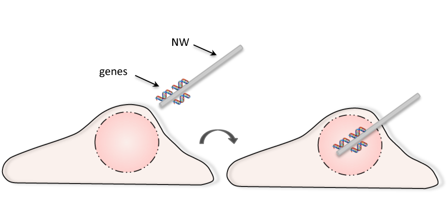
Nanowire-based endoscopy has attracted interest due to its ability to manipulate cells at the single-cell level with minimal cellular perturbation. High-density, vertically aligned nanowire arrays have been used as an efficient gene delivery system. Despite the high transfection rates, culturing the cells on nanowire arrays might have other influences on the cellular behaviour. For example, stem cells cultured on silicon nanowires show significantly different adhesion, proliferation and differentiation, compared with flat silicon or other control substrates. Furthermore, such arrays are not location-specific and require optimization of the nanowire density and dimension for the different the cell types. In collaboration with the group of Prof. Hiroshi Uji-i we are developing a method to delivery genetic material using a single nanowire. In contrast to the existing methods, this approach can be applied to any cell type and is extremely specific: it can target a single cell and it can deliver the genetic material exactly at the desired position, such as inside of the nucleus, with no damage to the cell. Since gene editing is a stochastic event occurring in only a fraction of the cells, the transfer of genetic material (or proteins) is of crucial importance in genome editing methods, where the nucleases must be efficiently delivered. The duration and magnitude of the nuclease expression are critical parameters for the level of both on-target and off-target nuclease activity. Additionally, the dose of donor template DNA is important to ensure efficient homologous recombination. The proposed method offers the possibility to deliver different molecules at different times, in synchronization with the cell cycle. The lab of Prof. Uji-i is one of the first (and few) groups worldwide to have developed and optimized a novel nanoscopic technique using 1D nanowires, with a diameter of less than 100 nm, for SERS endoscopic studies. It has been already proven by us that the thin diameter and 1D structure of the NW greatly reduces the damage induced to a live cell during probe insertion. Although designed for a different purpose, this nanoprobe is ideal as a starting point to develop a new NW-based gene delivery system.
This paper is about the number 1. The number 2 is left for future work.
Published in Journal of Non-Crystalline Solids, 2004
Download here
All in one: Exceptionally photostable, highly fluorescent, water‐soluble, and monofunctional perylene and terrylene dyes bearing reactive groups for covalent attachment to biomolecules have been synthesized (see picture). Single‐molecule enzyme tracking revealed that single enzymes could be visualized even on a substrate with fluorescent background.
Published in Angewandte Chemie, 2008
Download here
First article reporting my work.
Published in ChemPhysChem, 2009
Download here
Insight into the exciton dynamics occurring in a polyfluorene-perylenediimide (PF-PDI) copolymer with a reaction mixture ratio of 100 fluorene units to 1 N,N′-bis(phenyl)-1,6,7,12-tetra(p-tert-octylphenoxy)-perylene-3,4,9,10-tetracarboxylic acid diimide (PDI) is presented here. Time-correlated single photon counting and femtosecond transient absorption spectroscopy measurements on the PF-PDI copolymer have been employed to investigate the excited-state properties of the polyfluorene subunit where the exciton is localized (PF) and the incorporated PDI chromophore. The experimental results were compared with those obtained from a polyfluorene polymer (model PF) and a N,N′-bis(2,6-diisopropylphenyl)-1,6,7,12-tetra(p-tert-octylphenoxy)-perylene-3,4,9,10-tetracarboxylic acid diimide (model PDI) which were used as reference compounds. Because of the high polydispersity of the PF-PDI copolymer, there is a polymer fraction present that contains no PDI chromophores (polyfluorene polymer fraction (PF polymer fraction)), and wide-field imaging of single polymers chains of the synthesized PF-PDI copolymer was used to estimate this PF polymer fraction. Following the primary excitation of the PF in the PF-PDI copolymer, energy hopping between PF’s can occur. A fraction of the energy of the absorbed photons will be transferred to a PDI chromophore via energy transfer from a PF. In a polar solvent, a charge transfer state having the S1 of the PDI moiety as a precursor state is found to form with high efficiency on a nanosecond time scale. The data suggest that a fraction of the absorbed energy is directed, transferred, and used in charge separation, providing a clear view of a multistep mechanism of exciton dissociation into charges.
Published in The Journal of Physical Chemistry B, 2010
Download here
We monitored the action of phospholipase A2 (PLA2) on L- and D-dipalmitoyl-phosphatidylcholine (DPPC) Langmuir monolayers by mounting a Langmuir-trough on a wide-field fluorescence microscope with single molecule sensitivity. This made it possible to directly visualize the activity and diffusion behavior of single PLA2 molecules in a heterogeneous lipid environment during active hydrolysis. The experiments showed that enzyme molecules adsorbed and interacted almost exclusively with the fluid region of the DPPC monolayers. Domains of gel state L-DPPC were degraded exclusively from the gel-fluid interface where the buildup of negatively charged hydrolysis products, fatty acid salts, led to changes in the mobility of PLA2. The mobility of individual enzymes on the monolayers was characterized by single particle tracking. Diffusion coefficients of enzymes adsorbed to the fluid interface were between 3.2 μm2/s on the L-DPPC and 4.9 μm2/s on the D-DPPC monolayers. In regions enriched with hydrolysis products, the diffusion dropped to ≈0.2 μm2/s. In addition, slower normal and anomalous diffusion modes were seen at the L-DPPC gel domain boundaries where hydrolysis took place. The average residence times of the enzyme in the fluid regions of the monolayer and on the product domain were between ≈30 and 220 ms. At the gel domains it was below the experimental time resolution, i.e., enzymes were simply reflected from the gel domains back into solution.
Published in Biophysical journal, 2010
Download here
Accumulating evidence indicates that membrane lipids are not randomly distributed but rather form specific domains. In particular, raft-like microdomains composed of cholesterol and sphingolipids are attracting a lot of attention. These microdomains are thought to serve as platforms for signal transduction and molecular trafficking, but it is difficult to elucidate their detailed structure since their reported size is smaller than the resolution of light microscopy. To circumvent this limitation, we designed probes for cholesterol- and sphingolipid-enriched microdomains dedicated for superresolution microscopy, PALM. The probes utilise the affinity of the toxins, θ-toxin and lysenin, for the cholesterol- and sphingomyelin-enriched membranes, respectively. The toxicity can be avoided by using non-toxic domains that retain the specific binding to the aforementioned membranes. The probes can easily be produced in E. coli as recombinant protein domains of toxins fused to a photoswitchable fluorescent protein, Dronpa. PALM imaging with these probes revealed two types of cholesterol-enriched microdomains, line-shaped ones with widths of around 150 nm and round ones with an average radius of 118 nm. All sphingomyelin-enriched microdomains were round with an average radius of 124 nm. Both the cholesterol- and sphingomyelin-enriched microdomains vanished by the depletion of cholesterol. The sphingomyelin-enriched microdomains also vanished by the depletion of sphingomyelin whereas the cholesterol-enriched microdomains were unaffected. We conclude that cholesterol- and sphingomyelin-enriched domains occupy different regions on the plasma membrane.
Published in Chemical Science, 2011
Download here
Platelet-decorated von Willebrand factor (VWF) strings anchored to the endothelial surface are rapidly cleaved by ADAMTS13. Individual VWF string characteristics such as number, location, and auxiliary features of the ADAMTS13 cleavage sites were explored here using imaging and computing software. By following changes in VWF string length, we demonstrated that VWF strings are cleaved multiple times, successively shortening string length in the function of time and generating fragments ranging in size from 5 to over 100 μm. These are larger than generally observed in normal plasma, indicating that further proteolysis takes place in circulation. Interestingly, in 89% of all cleavage events, VWF strings elongate precisely at the cleavage site before ADAMTS13 proteolysis. These local elongations are a general characteristic of VWF strings, independent of the presence of ADAMTS13. Furthermore, large elongations, ranging in size from 1.4 to 40 μm, occur at different sites in space and time. In conclusion, ADAMTS13-mediated proteolysis of VWF strings under flow is preceded by large elongations of the string at the cleavage site. These elongations may lead to the simultaneous exposure of many exosites, thereby facilitating ADAMTS13-mediated cleavage.
Published in Journal of Biological Chemistry, 2011
Download here
Virus assembly and interaction with host-cell proteins occur at length scales below the diffraction limit of visible light. Novel super-resolution microscopy techniques achieve nanometer resolution of fluorescently labeled molecules. The cellular restriction factor tetherin (also known as CD317, BST-2 or HM1.24) inhibits the release of human immunodeficiency virus 1 (HIV-1) through direct incorporation into viral membranes and is counteracted by the HIV-1 protein Vpu. For super-resolution analysis of HIV-1 and tetherin interactions, we established fluorescence labeling of HIV-1 proteins and tetherin that preserved HIV-1 particle formation and Vpu-dependent restriction, respectively. Multicolor super-resolution microscopy revealed important structural features of individual HIV-1 virions, virus assembly sites and their interaction with tetherin at the plasma membrane. Tetherin localization to micro-domains was dependent on both tetherin membrane anchors. Tetherin clusters containing on average 4 to 7 tetherin dimers were visualized at HIV-1 assembly sites. Combined biochemical and super-resolution analysis revealed that extended tetherin dimers incorporate both N-termini into assembling virus particles and restrict HIV-1 release. Neither tetherin domains nor HIV-1 assembly sites showed enrichment of the raft marker GM1. Together, our super-resolution microscopy analysis of HIV-1 interactions with tetherin provides new insights into the mechanism of tetherin-mediated HIV-1 restriction and paves the way for future studies of virus-host interactions.
Published in PLoS pathogens, 2011
Download here
Photoactivation localization microscopy (PALM) was applied to study surface‐enhanced fluorescence (SEF) on metal nanostructures (SEF‐PALM). The detection of fluorescence from individual single molecules can be used to image the point‐spread‐function and spatial distribution of the fluorescence emitted in the vicinity of a metal surface. Due to the strong scattering effect, the angular distribution of the fluorescence is altered by metals, resulting in a spatial shift of fluorescence spots with respect to the metal nanostructures, and has to be taken into account in the analysis. SEF‐PALM can be used to discriminate effects of labelling density when estimating the enhancement factor in SEF. Furthermore, nanostructures with sizes below the diffraction limit can be resolved using this technique. SEF‐PALM is established as a powerful tool to study plasmon‐mediated phenomena on metal nanostructures.
Published in ChemPhysChem, 2012
Download here
An anionic fluorene-phenylene poly{1,4-phenylene-[9,9-bis(4-phenoxy-butylsulfonate)]fluorene-2,7-diyl}-based copolymer containing on-chain perylenediimine (PDI) chromophoric units, PBS-PFP-PDI, was synthesized and its photophysical properties studied as aggregates and isolated chains in water and dioxane/water (1:1) solution. UV–vis and emission spectroscopy measurements, time-correlated single photon counting, and wide field imaging have been employed to investigate the excited-state behavior of the PBS-PFP-PDI copolymer, including the effect of environment on the energy and electron transfer to the on-chain PDI chromophore. Although the Förster overlap integral is favorable, no evidence is found for intramolecular singlet excitation energy transfer in isolated copolymer chains in solution. Fluorescence is suggested to involve an interchain process, thus revealing that isolated copolymer chains in solution do not undergo efficient intramolecular energy transfer. However, quenching of the PBS-PFP excited state by PDI is observed in aqueous media and ultrafast pump–probe studies in water or dioxane–water solutions show that electron transfer occurs from the phenylene-fluorene units to the PDI. The extent of electron transfer increases with aggregation, suggesting it is largely an interchain process. The interaction of the negatively charged PBS-PFP-PDI copolymer with the positively charged surfactant hexadecyltrimethylammonium bromide (CTAB) in solution has also been studied. The copolymer PBS-PFP-PDI aggregates with the surfactant already at concentrations below the critical micelle concentration (cmc) and the nonpolar environment allows intermolecular energy transfer, observed by the weak emission band located at 630 nm that is associated with the emission of the PDI chromophore. However, the fact that the PDI photoluminescence (PL) lifetime (∼1.4 ns) obtained in the presence of CTAB is considerably shorter than that of the nonaggregated chromophore (∼5.4 ns) suggests that even in this case there is considerable PL quenching, possibly through some charge transfer route. The increase of the PBS-PFP-PDI photoluminescence intensity at surfactant concentrations above the cmc indicates deaggregation of polyelectrolyte within the initially formed polyelectrolyte–surfactant aggregates.
Published in The Journal of Physical Chemistry B, 2012
Download here
The spatio-temporal membrane behavior of glycine receptors (GlyRs) is known to be of influence on receptor homeostasis and functionality. In this work, an elaborate fluorimetric strategy was applied to study the GlyR α3K and L isoforms. Previously established differential clustering, desensitization and synaptic localization of these isoforms imply that membrane behavior is crucial in determining GlyR α3 physiology. Therefore diffusion and aggregation of homomeric α3 isoform-containing GlyRs were studied in HEK 293 cells. A unique combination of multiple diffraction-limited ensemble average methods and subdiffraction single particle techniques was used in order to achieve an integrated view of receptor properties. Static measurements of aggregation were performed with image correlation spectroscopy (ICS) and, single particle based, direct stochastic optical reconstruction microscopy (dSTORM). Receptor diffusion was measured by means of raster image correlation spectroscopy (RICS), temporal image correlation spectroscopy (TICS), fluorescence recovery after photobleaching (FRAP) and single particle tracking (SPT). The results show a significant difference in diffusion coefficient and cluster size between the isoforms. This reveals a positive correlation between desensitization and diffusion and disproves the notion that receptor aggregation is a universal mechanism for accelerated desensitization. The difference in diffusion coefficient between the clustering GlyR α3L and the non-clustering GlyR α3K cannot be explained by normal diffusion. SPT measurements indicate that the α3L receptors undergo transient trapping and directed motion, while the GlyR α3K displays mild hindered diffusion. These findings are suggestive of differential molecular interaction of the isoforms after incorporation in the membrane.
Published in Biochimica et Biophysica Acta (BBA)-Biomembranes, 2012
Download here
Vessel sprouting by migrating tip and proliferating stalk endothelial cells (ECs) is controlled by genetic signals (such as Notch), but it is unknown whether metabolism also regulates this process. Here, we show that ECs relied on glycolysis rather than on oxidative phosphorylation for ATP production and that loss of the glycolytic activator PFKFB3 in ECs impaired vessel formation. Mechanistically, PFKFB3 not only regulated EC proliferation but also controlled the formation of filopodia/lamellipodia and directional migration, in part by compartmentalizing with F-actin in motile protrusions. Mosaic in vitro and in vivo sprouting assays further revealed that PFKFB3 overexpression overruled the pro-stalk activity of Notch, whereas PFKFB3 deficiency impaired tip cell formation upon Notch blockade, implying that glycolysis regulates vessel branching.
Published in Cell, 2013
Download here
The gating of Ca2+-activated Cl− channels is controlled by a complex interplay among [Ca2+]i, membrane potential and permeant anions. Besides Ca2+, Ba2+ also can activate both TMEM16A and TMEM16B. This study reports the effects of several divalent cations as regulators of TMEM16A channels stably expressed in HEK293T cells. Among the divalent cations that activate TMEM16A, Ca2+ is most effective, followed by Sr2+ and Ni2+, which have similar affinity, while Mg2+ is ineffective. Zn2+ does not activate TMEM16A but inhibits the Ca2+-activated chloride currents. Maximally effective concentrations of Sr2+ and Ni2+ occluded activation of the TMEM16A current by Ca2+, which suggests that Ca2+, Sr2+ and Ni2+ all regulate the channel by the same mechanism.
Published in The Journal of Membrane Biology, 2013
Download here
The epidermal growth factor (EGF) receptor transduces the extracellular EGF signal into the cells. The distribution of these EGF receptors in the plasma membrane is heterogeneous and dynamic, which is proposed to be important for the regulation of cell signaling. The response of the cells to a physiological concentration of EGF is not homogeneous, which makes it difficult to analyze the dynamics related to the response. Here we developed a system to perform functional imaging during single particle tracking (SPT) analysis. This system made it possible to observe the cytosolic Ca2+ concentration to monitor the cell response while tracking individual EGF molecules and found that about half of the cells responded to the stimulation with 1.6 nM EGF. In the responding cells, the EGF receptor showed 3 modes of movement: fast (the diffusion coefficient of 0.081 ± 0.009 μm2/sec, 29 ± 9%), slow (0.020 ± 0.005 μm2/sec, 22 ± 6%), and stationary (49 ± 13%). The diffusion coefficient of the fast mode movement in the responding cells was significantly larger than that in the nonresponding cells (0.069 ± 0.009 μm2/sec, p < 0.05). The diffusion coefficient of the fast mode movement is thought to reflect the monomer–dimer equilibrium of the EGF receptor. We assumed that the feedback regulation via the Ca2+ signaling pathway slightly shifts the equilibrium from dimer to monomer in the responding cells.
Published in Biophysical Reviews and Letters, 2013
Download here
The ultrafast excited state dynamics of the fluorescent protein Kaede has been investigated by employing time resolved fluorescence and transient absorption. Upon irradiation of its neutral state, the protein undergoes an efficient conversion to a state that fluoresces at longer wavelengths. The molecular basis of the photoconversion involves an expansion of the chromophore π-conjugation by formal β-elimination but details of the reaction pathway remain subject to debate. Based on the kinetics observed in experiments on the protein sample in both H2O and D2O buffers, we suggest that a light-initiated cleavage mechanism (20 ps) could take place, forming the neutral red state in which the red chromophore resides. Excitation of the neutral red form results in the formation of the red anionic species via two Förster resonance energy transfer (FRET) channels. FRET between red neutral and red anionic forms occurs within the tetramer with time constants of 13.4 ps and 210 ps. In contrast to literature proposals no ESPT was observed
Published in Photochemical & Photobiological Sciences, 2013
Download here
In this study, we examined the intracellular whereabouts of Mrr, a cryptic type IV restriction endonuclease of Escherichia coli K12, in response to different conditions. In absence of stimuli triggering its activity, Mrr was found to be strongly associated with the nucleoid as a number of discrete foci, suggesting the presence of Mrr hotspots on the chromosome. Previously established elicitors of Mrr activity, such as exposure to high (hydrostatic) pressure (HP) or expression of the HhaII methyltransferase, both caused nucleoid condensation and an unexpected coalescence of Mrr foci. However, although the resulting Mrr/nucleoid complex was stable when triggered with HhaII, it tended to be only short-lived when elicited with HP. Moreover, HP-mediated activation of Mrr typically led to cellular blebbing, suggesting a link between chromosome and cellular integrity. Interestingly, Mrr variants could be isolated that were specifically compromised in either HhaII- or HP-dependent activation, underscoring a mechanistic difference in the way both triggers activate Mrr. In general, our results reveal that Mrr can take part in complex spatial distributions on the nucleoid and can be engaged in distinct modes of activity.
Published in Nucleic Acids Research, 2014
Download here
Advanced imaging techniques crucially depend on the labels used. In this work, we present the structure-guided design of a fluorescent protein that displays both reversibly photochromic and green-to-red photoconversion behavior. We first designed ffDronpa, a mutant of the photochromic fluorescent protein Dronpa that matures up to three times faster while retaining its interesting photochromic features. Using a combined evolutionary and structure-driven rational design strategy, we developed a green-to-red photoconvertible ffDronpa mutant, called pcDronpa, and explored different optimization strategies that resulted in its improved version, pcDronpa2. This fluorescent probe combines a high brightness with low photobleaching and photoblinking. We herein show that, despite its tetrameric nature, pcDronpa2 allows for multimodal subdiffraction imaging by sequentially imaging a given sample using both super-resolution fluctuation imaging and localization microscopy.
Published in ACS Nano, 2014
Download here
Developing molecular systems with functions analogous to those of macroscopic machine components, such as rotors, gyroscopes and valves, is a long-standing goal of nanotechnology. However, macroscopic analogies go only so far in predicting function in nanoscale environments, where friction dominates over inertia. In some instances, ratchet mechanisms have been used to bias the ever-present random, thermally driven (Brownian) motion and drive molecular diffusion in desired directions. Here, we visualize the motions of surface-bound molecular rotors using defocused fluorescence imaging, and observe the transition from hindered to free Brownian rotation by tuning medium viscosity. We show that the otherwise random rotations can be biased by the polarization of the excitation light field, even though the associated optical torque is insufficient to overcome thermal fluctuations. The biased rotation is attributed instead to a fluctuating-friction mechanism in which photoexcitation of the rotor strongly inhibits its diffusion rate.
Published in Nature Nanotechnology, 2014
Download here
von Willebrand factor (VWF) strings are removed from the endothelial surface by ADAMTS13 (a disintegrin and metalloprotease with thrombospondin type-1 repeats)-mediated proteolysis. To visualize how single ADAMTS13 molecules bind to these long strings, we built a customized single molecule fluorescence microscope and developed single particle tracking software. Extensive analysis of over 6,000 single inactive ADAMTS13E225Q enzymes demonstrated that 20% of these molecules could be detected in at least two consecutive 60-ms frames and followed two types of trajectories. ADAMTS13E225Q molecules either decelerated in the vicinity of VWF strings, whereas sometimes making brief contact with the VWF string before disappearing again, or readily bound to the VWF strings and this for 120 ms or longer. These interactions were observed at several sites along the strings. Control experiments using an IgG protein revealed that only the second type of trajectory reflected a specific interaction of ADAMTS13 with the VWF string. In conclusion, we developed a dedicated single molecule fluorescence microscope for detecting single ADAMTS13 molecules (nm scale) on their long, flow-stretched VWF substrates (μm scale) anchored on living cells. Comprehensive analysis of all detected enzymes showed a random interaction mechanism for ADAMTS13 with many available binding sites on the VWF strings.
Published in Journal of Biological Chemistry, 2014
Download here
Single particle tracking (SPT) of transmembrane receptors in the plasma membrane often reveals heterogeneous diffusion. A thorough interpretation of the displacements requires an extensive analysis suited for discrimination of different motion types present in the data. Here the diffusion pattern of the homomeric α3-containing glycine receptor (GlyR) is analyzed in the membrane of HEK 293 cells. More specifically, the influence of the α3 RNA splice variants α3K and α3L on lateral membrane diffusion of the receptor is revealed in detail. Using a combination of ensemble and local SPT analysis, free and anomalous diffusion parameters are determined. The GlyR α3 free diffusion coefficient is found to be 0.13 ± 0.01 μm2/s and both receptor variants display confined motion. The confinement probability level and residence time are significantly elevated for the α3L variant compared to the α3K variant. Furthermore, for the α3L GlyR, the presence of directed motion was also established, with a velocity matching that of saltatory vesicular transport. These findings reveal that α3 GlyRs are prone to different types of anomalous diffusion and reinforce the role of RNA splicing in determining lateral membrane trafficking.
Published in Biochimica et Biophysica Acta (BBA)-Molecular Cell Research, 2014
Download here
Employing viruses as nanoscopic lipid-enveloped test tubes allows the miniaturization of protein–protein interaction (PPI) assays while preserving the physiological environment necessary for particular biological processes. Applied to the study of the human immunodeficiency virus type 1 (HIV-1), viral biology and pathology can also be investigated in novel ways, both in vitro as well as in infected cells. In this work we report on an experimental strategy that makes use of engineered HIV-1 viral particles, to allow for probing PPIs of the HIV-1 integrase (IN) inside viruses with single-molecule Förster resonance energy transfer (FRET) using fluorescent proteins (FP). We show that infectious fluorescently labeled viruses can be obtained and that the quantity of labels can be accurately measured and controlled inside individual viral particles. We demonstrate, with proper control experiments, the formation of IN oligomers in single viral particles and inside viral complexes in infected cells. Finally, we show a clear effect on IN oligomerization of small molecule inhibitors of interactions of IN with its natural human cofactor LEDGF/p75, corroborating that IN oligomer enhancing drugs are active already at the level of the virus and strongly suggesting the presence of a dynamic, enhanceable equilibrium between the IN dimer and tetramer in viral particles. Although applied to the HIV-1 IN enzyme, our methodology for utilizing HIV virions as nanoscopic test tubes for probing PPIs is generic, i.e., other PPIs targeted into the HIV-1, or PPIs targeted into other viruses, can potentially be studied with a similar strategy.
Published in ACS Nano, 2014
Download here
Important cellular events such as division require drastic changes in the shape of the membrane. These remodeling processes can be triggered by the binding of specific proteins or by changes in membrane composition and are linked to phospholipid metabolism for which dedicated enzymes, named phospholipases, are responsible. Here wide-field fluorescence microscopy is used to visualize shape changes induced by the action of phospholipase A1 on dye-labeled supported membranes of POPC (1-palmitoyl-2-oleoly-sn-glycero-3-phosphocholine). Time-lapse imaging demonstrates that layers either shrink and disappear or fold and collapse into vesicles. These vesicles can undergo further transformations such as budding, tubulation, and pearling within 5 min of formation. Using dye-labeled phospholipases, we can monitor the presence of the enzyme at specific positions on the membrane as the shape transformations occur. Furthermore, incorporating the products of hydrolysis into POPC membranes is shown to induce transformations similar to those observed for enzyme action. The results suggest that phospholipase-mediated hydrolysis plays an important role in membrane transformations by altering the membrane composition, and a model is proposed for membrane curvature based on the presence and shape of hydrolysis products.
Published in Langmuir, 2014
Download here
In this study, the effect of glycine receptor (GlyR) α3 alternative RNA splicing on the distribution of receptors in the membrane of human embryonic kidney 293 cells is investigated using optical super-resolution microscopy. Direct stochastic optical reconstruction microscopy is used to image both α3K and α3L splice variants individually and together using single- and dual-color imaging. Pair correlation analysis is used to extract quantitative measures from the resulting images. Autocorrelation analysis of the individually expressed variants reveals clustering of both variants, yet with differing properties. The cluster size is increased for α3L compared to α3K (mean radius 92 ± 4 and 56 ± 3 nm, respectively), yet an even bigger difference is found in the cluster density (9,870 ± 1,433 and 1,747 ± 200 μm−2, respectively). Furthermore, cross-correlation analysis revealed that upon co-expression, clusters colocalize on the same spatial scales as for individually expressed receptors (mean co-cluster radius 94 ± 6 nm). These results demonstrate that RNA splicing determines GlyR α3 membrane distribution, which has consequences for neuronal GlyR physiology and function.
Published in Histochemistry and Cell Biology, 2014
Download here
Live‐cell surface‐enhanced Raman spectroscopy (SERS) endoscopy is developed by using plasmonic nanowire waveguides as endoscopic probes. It is demonstrated that the probe insertion does not stress the cell. Opposed to conventional SERS endoscopy, with excitation at the hotspot within the cell, the remote excitation method yields low‐background SERS spectra from specific cell compartments with minimal associated photodamage.
Published in Advanced Materials, 2014
Download here
A new strategy has been designed for visualized detection of the conformation changes of calmodulin bound to target peptide (CaM-M13) based on the conformation sensitive property of a water-soluble conjugated polythiophene derivative (PMNT) and the electrostatic interactions of PMNT/CaM-M13. Interestingly, the direct visualized PMNT color changes under UV irradiation and the turbidity changes of samples in aqueous medium can be applied to detect the conformation changes as well as the controllable assembly of PMNT/CaM-M13 with Ca2+ in aqueous medium. Because of the specific binding of Ca2+, the assembly of PMNT/CaM-M13 can be applied to sense calcium as well.
Published in ACS Appl. Mater. Interfaces, 2014
Download here
Transportin-SR2 (Tnpo3, TRN-SR2), a human karyopherin encoded by the TNPO3 gene, has been identified as a cellular cofactor of HIV-1 replication, specifically interacting with HIV-1 integrase (IN). Whether this interaction mediates the nuclear import of HIV remains controversial. We previously characterized the TRN-SR2 binding interface in IN and introduced mutations at these positions to corroborate the biological relevance of the interaction. The pleiotropic nature of IN mutations complicated the interpretation. Indeed, all previously tested IN interaction mutants also affected reverse transcription (RT). Here we report on a virus with a pair of IN mutations, INR263A/K264A, that significantly reduce interaction with TRN-SR2. The virus retains wild-type reverse transcription activity but displays a block in nuclear import and integration, as measured by Q-PCR. The defect in integration of this mutant resulted in a smaller increase in the number of 2-LTR circles than for virus specifically blocked at integration by raltegravir or catalytic site mutations (IND64N/D116N/E152Q). Finally, using an eGFP-IN labeled HIV fluorescence-based import assay, the defect in nuclear import was corroborated. These data altogether underscore the importance of the HIV-IN TRN-SR2 protein-protein interaction for HIV nuclear import and validate the IN/TRN-SR2 interaction interface as a promising target for future antiviral therapy.
Published in Journal of Biological Chemistry, 2014
Download here
The conformation of calmodulin (CaM) changes from closed configuration to open one, converting to a claviform dumbbell‐shaped biomolecule upon Ca2+‐binding. A hybrid probe of graphene oxide (GO) cationic conjugated polymer for detection of the conformation transition of CaM by using FRET technique is demonstrated. The stronger hydrophobic interaction and weaker electrostatic repulsion leads to more CaM adsorption to the surface of GO upon binding with Ca2+ than that of CaM in the absence of Ca2+ (apoCaM), resulting in much farther proximity between poly[(9,9‐bis(6′‐N,N,N‐trimethylammonium)hexyl)‐fluorenylene phenylene dibromide] (PFP) and green fluorescent protein labeled at the N‐terminus of CaM and therefore much weaker FRET efficiency for PFP/Ca2+/CaM in comparison with that of PFP/apoCaM in the presence of GO. Notably, the assembly of CaM with GO is quantitatively and reversibly controlled by Ca2+ ions.
Published in Adv. Funct.Mater., 2015
Download here
Photoactivated localization microscopy (PALM) is a super-resolution imaging technique based on the detection and subsequent localization of single fluorescent molecules. PALM is therefore a powerful tool in resolving structures and putative interactions of biomolecules at the ultimate analytical detection limit. However, its limited imaging depth restricts PALM mostly to in vitro applications. Considering the additional need for anatomical context when imaging a multicellular organism, these limitations render the use of PALM in whole animals difficult. Here we integrated PALM with confocal microscopy for correlated imaging of the C. elegans nervous system, a technique we termed confocal correlated PALM (ccPALM). The neurons, lying below several tissue layers, could be visualized up to 10 μm deep inside the animal. By ccPALM, we visualized ionotropic glutamate receptor distributions in C. elegans with an accuracy of 20 nm, revealing super-resolution structure of receptor clusters that we mapped onto annotated neurons in the animal. Pivotal to our results was the TIRF-independent detection of single molecules, achieved by genetic regulation of labeled receptor expression and localization to effectively reduce the background fluorescence. By correlating PALM with confocal microscopy, this platform enables dissecting biological structures with single molecule resolution in the physiologically relevant context of whole animals.
Published in Scientific Reports, 2015
Download here
The enhancement of molecular absorption, emission and scattering processes by coupling to surface plasmon polaritons on metallic nanoparticles is a key issue in plasmonics for applications in (bio)chemical sensing, light harvesting and photocatalysis. Nevertheless, the point spread functions for single-molecule emission near metallic nanoparticles remain difficult to characterize due to fluorophore photodegradation, background emission and scattering from the plasmonic structure. Here we overcome this problem by exciting fluorophores remotely using plasmons propagating along metallic nanowires. The experiments reveal a complex array of single-molecule fluorescence point spread functions that depend not only on nanowire dimensions but also on the position and orientation of the molecular transition dipole. This work has consequences for both single-molecule regime-sensing and super-resolution imaging involving metallic nanoparticles and opens the possibilities for fast size sorting of metallic nanoparticles, and for predicting molecular orientation and binding position on metallic nanoparticles via far-field optical imaging.
Published in Nature Communications, 2015
Download here
LSSmOrange is a fluorescent protein with a large energy gap between the absorption and emission bands (5275 cm–1). The electronic structure of the LSSmOrange chromophore, 2-[(5-)-2-hydroxy-dihydrooxazole]-4-(p-hydroxybenzylidene)-5-imidazolinone, is affected by deprotonation of the p-hydroxybenzylidene group. We investigated LSSmOrange by time-resolved spectroscopy in the femtosecond and nanosecond range. The ground state chromophore was almost exclusively in the neutral form, which had a main absorption band at 437 nm with a small shoulder at 475 nm. The absorption at a wavelength within the former band promoted the protein to the excited state where excited state proton transfer (ESPT) could lead to deprotonation in 0.8 ps. Following ESPT, the chromophore emitted fluorescence with a maximum at 573 nm and a decay time of 3500 ps. Although deprotonation by ESPT occurs, we unexpectedly found a slow accumulation of the anionic form in the ground state upon repeated high intensity excitation. This accumulation of the anionic form was accompanied by a shift of the absorption band to 553 nm without changing the emission band. MALDI-MS revealed that this shift is accompanied by decarboxylation of E222, which is interacting with the imidazolinone ring of the chromophore. We concluded that the photoinduced decarboxylation induced a conformational change that affected local environment around the hydroxyl group, resulting in a stable deprotonated form of the chromophore.
Published in The Journal of Physical Chemistry B, 2015
Download here
Optical antennas made of metallic nanostructures dramatically enhance single-molecule fluorescence to boost the detection sensitivity. Moreover, emission properties detected at the optical far field are dictated by the antenna. Here we study the emission from molecule–antenna hybrids by means of super-resolution localization and defocused imaging. Whereas gold nanorods make single-crystal violet molecules in the tip’s vicinity visible in fluorescence, super-resolution localization on the enhanced molecular fluorescence reveals geometrical centers of the nanorod antenna instead. Furthermore, emission angular distributions of dyes linked to the nanorod surface resemble that of nanorods in defocused imaging. The experimental observations are consistent with numerical calculations using the finite-difference time-domain method.
Published in ACS nano, 2016
Download here
Detection of carbon dioxide (CO2) is of fundamental importance in diverse applications ranging from environmental analysis to agricultural production. In this work, a hybrid probe based on guanidinium-pendent oligofluorene (G-OF) and water-soluble conjugated polythiophene (PTP) has been developed for the turn on detection of CO2 with low background signal, taking advantage of the efficient fluorescence quenching of the tight aggregate of G-OF/PTP. In the presence of CO2, the electrostatic repulsion between G-OF and PTP can be effectively enhanced through protonation of the side chains, leading to the disaggregation and thus the “turn-on” fluorescence. The strategy allows for the light-up visible detection of CO2 with high sensitivity. Importantly, this system is capable of sensitively monitoring the concentration changes of CO2 in the process of the photosynthesis, which represents a concept to monitor the photosynthesis based on water-soluble conjugated polymers.
Published in Anal. Chem., 2016
Download here
Nuclear entry is a selective, dynamic process granting the HIV-1 pre-integration complex (PIC) access to the chromatin. Classical analysis of nuclear entry of heterogeneous viral particles only yields averaged information. We now have employed single-virus fluorescence methods to follow the fate of single viral pre-integration complexes (PICs) during infection by visualizing HIV-1 integrase (IN). Nuclear entry is associated with a reduction in the number of IN molecules in the complexes while the interaction with LEDGF/p75 enhances IN oligomerization in the nucleus. Addition of LEDGINs, small molecule inhibitors of the IN-LEDGF/p75 interaction, during virus production, prematurely stabilizes a higher-order IN multimeric state, resulting in stable IN multimers resistant to a reduction in IN content and defective for nuclear entry. This suggests that a stringent size restriction determines nuclear pore entry. Taken together, this work demonstrates the power of single-virus imaging providing crucial insights in HIV replication and enabling mechanism-of-action studies.
Published in Scientific reports, 2016
Download here
Photoswitchable fluorescent proteins are capable of changing their spectral properties upon light irradiation, thus allowing one to follow a chosen subpopulation of molecules in a biological system. Recently, we revealed a photoinduced absorption band shift of LSSmOrange, which was originally engineered to have a large energy gap between excitation and emission bands. Here, we evaluated the performance of LSSmOrange as a fluorescent tracer in living cells. The absorption maximum of LSSmOrange in HeLa cells shifted from 437 nm to 553 nm upon illumination with a 405-, 445-, 458-, or 488-nm laser on a laser-scanning microscope, whereas the emission band remained same (∼570 nm). LSSmOrange behaves as a freely diffusing protein in living cells, enabling the use of the protein as a fluorescence tag for studies of protein dynamics. By targeting LSSmOrange in mitochondria, we observed an exchange of soluble molecules between the matrices upon mitochondrial fusion. Since converted and unconverted LSSmOrange proteins have similar emission spectra, this tracer offers unique possibilities for multicolor imaging. The fluorescence emission from LSSmOrange was spectrally distinguishable from that of eYFP and mRFP, and could be separated completely by applying linear unmixing. Furthermore, by using a femtosecond laser at 850 nm, we showed that a two-photon process could evoke a light-induced red shift of the absorption band of LSSmOrange, providing a strict confinement of the conversion volume in a three-dimensional space.
Published in Biophysical journal, 2016
Download here
The CO2‐responsive and biocatalytic assembly based on conjugated polymers has been demonstrated by combining the signal amplification property of the polythiophene derivative (PTP) and the catalytic actions of carbonic anhydrase (CA). CO2 is applied as a new trigger mode to construct the smart assembly by controlling the electrostatic and hydrophobic interactions between the PTP molecules in aqueous solution, leading to the visible fluorescence changes. Importantly, the assembly transformation of PTP can be specifically and highly accelerated by CA based on the efficient catalytic activity of CA for the inter‐conversion between CO2 and HCO3-, mimicking the CO2‐associated biological processes that occurred naturally in living organisms. Moreover, the PTP‐based assembly can be applied for biomimetic CO2 sequestration with fluorescence monitoring in the presence of CA and calcium.
Published in Macromol. Rapid Commun., 2017
Download here
Loss of ER Ca2+ homeostasis triggers endoplasmic reticulum (ER) stress and drives ER-PM contact sites formation in order to refill ER-luminal Ca2+. Recent studies suggest that the ER stress sensor and mediator of the unfolded protein response (UPR) PERK regulates intracellular Ca2+ fluxes, but the mechanisms remain elusive. Here, using proximity-dependent biotin identification (BioID), we identified the actin-binding protein Filamin A (FLNA) as a key PERK interactor. Cells lacking PERK accumulate F-actin at the cell edges and display reduced ER-PM contacts. Following ER-Ca2+ store depletion, the PERK-FLNA interaction drives the expansion of ER-PM juxtapositions by regulating F-actin-assisted relocation of the ER-associated tethering proteins Stromal Interaction Molecule 1 (STIM1) and Extended Synaptotagmin-1 (E-Syt1) to the PM. Cytosolic Ca2+ elevation elicits rapid and UPR-independent PERK dimerization, which enforces PERK-FLNA-mediated ER-PM juxtapositions. Collectively, our data unravel an unprecedented role of PERK in the regulation of ER-PM appositions through the modulation of the actin cytoskeleton.
Published in Molecular Cell, 2017
Download here
Enhancing the thermal stability of proteins is an important task for protein engineering. There are several ways to increase the thermal stability of proteins in biology, such as greater hydrophobic interactions, increased helical content, decreased occurrence of thermolabile residues, or stable hydrogen bonds. Here, we describe a well-defined polymer based on β-helical polyisocyanotripeptides (TriPIC) that uses biological approaches, including hydrogen bonding and hydrophobic interactions for its exceptional thermal stability in aqueous solutions. The multiple hydrogen bonding arrays along the polymer backbone shield the hydrophobic core from water. Variable temperature CD and FTIR studies indicate that, on heating, a better packed polymer conformation further stiffens the backbone. Driven by hydrophobic interactions, TriPIC solutions give fully reversible hydrogels that can withstand high temperatures (80 °C) for extended times. Cryo-scanning electron microscopy (cryo-SEM), small-angle X-ray scattering (SAXS), and thorough rheological analysis show that the hydrogel has a bundled architecture, which gives rise to strain stiffening effects on deformation of the gel, analogous to many biological hydrogels.
Published in Macromolecules, 2017
Download here
Correlative imaging by fluorescence and force microscopy is an emerging technology to acquire orthogonal information at the nanoscale. Whereas atomic force microscopy excels at resolving the envelope structure of nanoscale specimens, fluorescence microscopy can detect specific molecular labels, which enables the unambiguous recognition of molecules in a complex assembly. Whereas correlative imaging at the micrometer scale has been established, it remains challenging to push the technology to the single-molecule level. Here, we used an integrated setup to systematically evaluate the factors that influence the quality of correlative fluorescence and force microscopy. Optimized data processing to ensure accurate drift correction and high localization precision results in image registration accuracies of ∼25 nm on organic fluorophores, which represents a 2-fold improvement over the state of the art in correlative fluorescence and force microscopy. Furthermore, we could extend the Atto532 fluorophore bleaching time ∼2-fold, by chemical modification of the supporting mica surface. In turn, this enables probing the composition of macromolecular complexes by stepwise photobleaching with high confidence. We demonstrate the performance of our method by resolving the stoichiometry of molecular subpopulations in a heterogeneous EcoRV–DNA nucleoprotein ensemble.
Published in ACS nano, 2017
Download here
Conjugated polymers have been widely studied as flexible, versatile semiconductors in organic electronics. However, the material stability is one of the problems limiting their applications. Thus, understanding the degradation process of conjugated polymers is crucial. In this work, we monitored the chain scission of the model polymer MEH-PPV in chloroform solutions under different conditions by assessing its molecular weight using gel permeation chromatography and optical spectral measurements. We showed that changes in the UV-VIS spectrum can be seen only when the degradation has already progressed substantially. The fluorescence spectrum was found to be almost totally insensitive to the degradation stage of the polymers. We demonstrate that chain scission in solutions happens even in the dark leading to a 15% decrease of the molecular weight after just one day of storage. If exposed to room light, the chain length decreases by about 10 times over one day of exposure. Using stronger light intensity or enriching the solution with oxygen accelerates the degradation process dramatically. The rate of the reaction follows approximately a square root dependence with light intensity and oxygen concentration. We conclude that some extent of polymer degradation is difficult to avoid in common laboratory practices since to prevent it, one needs to work in an oxygen-free atmosphere in the dark. Preparation of polymer films from partially degraded solutions might lead not only to losing the connection between the molecular weight and the opto-electronic properties but also to unintentional doping of the semiconductor by products of chain scission reactions.
Published in Physical Chemistry Chemical Physics, 2018
Download here
Expression cloning from cDNA is an important technique for acquiring genes encoding novel fluorescent proteins. However, the probability of in-frame cDNA insertion following the first start codon of the vector is normally only 1/3, which is a cause of low cloning efficiency. To overcome this issue, we developed a new expression plasmid vector, pRSET-TriEX, in which transcriptional slippage was induced by introducing a DNA sequence of (dT)14 next to the first start codon of pRSET. The effectiveness of frame-insensitive cloning was validated by inserting the gene encoding eGFP with all three possible frames to the vector. After transformation with one of these plasmids, E. coli cells expressed eGFP with no significant difference in the expression level. The pRSET-TriEX vector was then used for expression cloning of a novel fluorescent protein from Scolionema suvaense. We screened 3658 E. coli colonies transformed with pRSET-TriEX containing Scolionema suvaense cDNA, and found one colony expressing a novel green fluorescent protein, ScSuFP. The highest score in protein sequence similarity was 42% with the chain c of multi-domain green fluorescent protein like protein “ember” from Anthoathecata sp. Variations in the N- and/or C-terminal sequence of ScSuFP compared to other fluorescent proteins indicate that the expression cloning, rather than the sequence similarity-based methods, was crucial for acquiring the gene encoding ScSuFP. The absorption maximum was at 498 nm, with an extinction efficiency of 1.17 × 10⁵ M−1·cm−1. The emission maximum was at 511 nm and the fluorescence quantum yield was determined to be 0.6. Pseudo-native gel electrophoresis showed that the protein forms obligatory homodimers.
Published in International journal of molecular sciences, 2018
Download here
Localization‐based super‐resolution microscopy has become an indispensable tool in biology to study features smaller than the diffraction limit of light. In a multicolor approach, adequate spectral separation of the different photoswitchable probes is required. A far‐red emitting dye is often one of the labels of choice. However, irradiation with high laser intensity can induce photo‐conversion of some of the most frequently used fluorophores. Herein we show that upon intense irradiation with a 561 nm laser line, far‐red organic dyes photoconvert to blue‐shifted emissive species. In the case of Alexa Fluor 647, the most commonly used fluorescent label in super‐resolution microscopy, this derivative is created over time in an intramolecular, irreversible photoinduced chemical reaction. The dynamics of this reaction are altered by the presence of reducing agents. Importantly, the blue‐shifted derivatives emit in the spectral range of the red fluorescent proteins (e. g. PAmCherry and converted mEos3.2), severely implicating multicolor super‐resolution imaging.
Published in ChemPhotoChem, 2018
Download here
Viruses are simple agents exhibiting complex reproductive mechanisms. Decades of research have provided crucial basic insights, antiviral medication and moderately successful gene therapy trials. The most infectious viral particle is, however, not always the most abundant one in a population, questioning the utility of classic ensemble-averaging virology. Indeed, viral replication is often not particularly efficient, prone to errors or containing parallel routes. Here, we review different single-molecule sensitive fluorescence methods that we employ routinely to investigate viruses. We provide a brief overview of the microscopy hardware needed and discuss the different methods and their application. In particular, we review how we applied (i) single-molecule Förster resonance energy transfer (smFRET) to probe the subviral human immunodeficiency virus (HIV-1) integrase (IN) quaternary structure;(ii) single particle tracking to study interactions of the simian virus 40 with membranes;(iii) 3D confocal microscopy and smFRET to quantify the HIV-1 pre-integration complex content and quaternary structure;(iv) image correlation spectroscopy to quantify the cytosolic HIV-1 Gag assembly, and finally;(v) super-resolution microscopy to characterize the interaction of HIV-1 with tetherin during assembly. We hope this review is an incentive for setting up and applying similar single-virus imaging studies in daily virology practice.
Published in Viruses, 2018
Download here
Protein–protein interactions (PPIs) form the basis of cellular processes, regulating cell behavior and fate. PPIs can be extremely transient in nature, which hinders their detection. In addition, traditional biochemical methods provided limited information on the spatial distribution and temporal dynamics of PPIs that is crucial for their regulation in the crowded cellular environment. Given the pivotal role of membrane micro- and nanodomains in the regulation of PPIs at the plasma membrane, the development of methods to visualize PPIs with a high spatial resolution is imperative. Here, we present a super-resolution fluorescence microscopy technique that can detect and map short-lived transient protein–protein interactions on a nanometer scale in the cellular environment. This imaging method is based on single-molecule fluorescence microscopy and exploits the effect of the difference in the mobility between cytosolic and membrane-bound proteins in the recorded fluorescence signals. After the development of the proof of concept using a model system based on membrane-bound modular protein domains and fluorescently labeled peptides, we applied this imaging approach to investigate the interactions of cytosolic proteins involved in the epidermal growth factor signaling pathway (namely, Grb2, c-Raf, and PLCγ1). The detected clusters of Grb2 and c-Raf were correlated with the distribution of the receptor at the plasma membrane. Additionally, the interactions of wild type PLCγ1 were compared with those detected with truncated mutants, which provided important information regarding the role played by specific domains in the interaction with the membrane. The results presented here demonstrate the potential of this technique to unravel the role of membrane heterogeneity in the spatiotemporal regulation of cell signaling.
Published in ACS Nano, 2018
Download here
Glutamine synthetase, encoded by the gene GLUL, is an enzyme that converts glutamate and ammonia to glutamine. It is expressed by endothelial cells, but surprisingly shows negligible glutamine-synthesizing activity in these cells at physiological glutamine levels. Here we show in mice that genetic deletion of Glul in endothelial cells impairs vessel sprouting during vascular development, whereas pharmacological blockade of glutamine synthetase suppresses angiogenesis in ocular and inflammatory skin disease while only minimally affecting healthy adult quiescent endothelial cells. This relies on the inhibition of endothelial cell migration but not proliferation. Mechanistically we show that in human umbilical vein endothelial cells GLUL knockdown reduces membrane localization and activation of the GTPase RHOJ while activating other Rho GTPases and Rho kinase, thereby inducing actin stress fibres and impeding endothelial cell motility. Inhibition of Rho kinase rescues the defect in endothelial cell migration that is induced by GLUL knockdown. Notably, glutamine synthetase palmitoylates itself and interacts with RHOJ to sustain RHOJ palmitoylation, membrane localization and activation. These findings reveal that, in addition to the known formation of glutamine, the enzyme glutamine synthetase shows unknown activity in endothelial cell migration during pathological angiogenesis through RHOJ palmitoylation.
Published in Nature, 2018
Download here
Mixed micelles based on amphiphilic gadolinium(III)-DOTA and europium(III)-DTPA complexes were synthesized and evaluated for their paramagnetic and optical properties as potential bimodal contrast agents. Amphiphilic folate molecule for targeting the folate receptor protein, which is commonly expressed on the surface of many human cancer cells, was used in the self-assembly process in order to create nanoaggregates with targeting properties. Both targeted and nontargeted nanoaggregates formed monodisperse micelles having distribution maxima of 10 nm. The micelles show characteristic europium(III) emission with quantum yields of 2% and 1.1% for the nontargeted and targeted micelles, respectively. Fluorescence microscopy using excitation at 405 nm and emission at 575–675 nm was employed to visualize the nanoaggregates in cultured HeLa cells. The uptake of folate-targeted and nontargeted micelles is already visible after 5 h of incubation and was characterized with the europium(III) emission, which is clearly observable in the cytoplasm of the cells. The very fast longitudinal relaxivity r1 of ca. 26 s–1 mM–1 per gadolinium(III) ion was observed for both micelles at 60 MHz and 310 K. Upon increasing the magnetic field to 300 MHz, the nanoaggregates exhibited a large switching to transversal relaxivity with r2 value of ca. 52 s–1 mM–1 at 310 K. Theoretical fitting of the 1H NMRD profiles indicate that the efficient T1 and T2 relaxations are sustained by the favorable magnetic and electron-configuration properties of the gadolinium(III) ion, rotational correlation time, and coordinated water molecule. These nanoaggregates could have versatile application as a positive contrast agent at the currently used magnetic imaging field strengths and a negative contrast agent in higher field applications, while at the same time offering the possibility for the loading of hydrophobic therapeutics or targeting molecules.
Published in ACS Appl. Mater. Interfaces, 2019
Download here
One of the promises of synthetic materials in cell culturing is that control over their molecular structures may ultimately be used to control their biological processes. Synthetic polymer hydrogels from polyisocyanides (PIC) are a new class of minimal synthetic biomaterials for three-dimensional cell culturing. The macromolecular lengths and densities of biofunctional groups that decorate the polymer can be readily manipulated while preserving the intrinsic nonlinear mechanics, a feature commonly displayed by fibrous biological networks. In this work, we propose the use of PIC gels as cell culture platforms with decoupled mechanical inputs and biological cues. For this purpose, different types of cells were encapsulated in PIC gels of tailored compositions that systematically vary in adhesive peptide (GRGDS) density, polymer length, and concentration; with the last two parameters controlling the gel mechanics. Both cancer and smooth muscle cells grew into multicellular spheroids with proliferation rates that depend on the adhesive GRGDS density, regardless of the polymer length, suggesting that for these cells, the biological input prevails over the mechanical cues. In contrast, human adipose-derived stem cells do not form spheroids but rather spread out. We find that the morphological changes strongly depend on the adhesive ligand density and the network mechanics; gels with the highest GRGDS densities and the strongest stiffening response to stress show the strongest spreading. Our results highlight the role of the nonlinear mechanics of the extracellular matrix and its synthetic mimics in the regulation of cell functions.
Published in Biomacromolecules, 2019
Download here
The pituitary is the master endocrine gland, harboring stem cells of which the phenotype and role remain poorly characterized. Here, we established organoids from mouse pituitary with the aim to generate a novel research model to study pituitary stem cell biology. The organoids originated from the pituitary cells expressing the stem cell marker SOX2 were long-term expandable, displayed a stemness phenotype during expansive culture and showed specific hormonal differentiation ability, although limited, after subrenal transplantation. Application of the protocol to transgenically injured pituitary harboring an activated stem cell population, resulted in more numerous organoids. Intriguingly, these organoids presented with a cystic morphology, whereas the organoids from undamaged gland were predominantly dense and appeared more limited in expandability. Transcriptomic analysis revealed distinct epithelial phenotypes and showed that cystic organoids more resembled the pituitary phenotype, at least to an immature state, and displayed in vitro differentiation, although yet moderate. Organoid characterization further exposed facets of regulatory pathways of the putative stem cells of the pituitary and advanced new injury-activated markers. Taken together, we established a novel organoid research model revealing new insights into the identity and regulation of the putative pituitary stem cells. This organoid model may eventually lead to an interesting tool to decipher pituitary stem cell biology in both healthy and diseased gland.
Published in Journal of Endocrinology, 2019
Download here
Most targeting strategies of anticancer drug delivery systems (DDSs) rely on the surface functionalization of nanocarriers with specific ligands, which trigger the internalization in cancer cells via receptor-mediated endocytosis. The endocytosis implies the entrapment of DDSs in acidic vesicles (endosomes and lysosomes) and their eventual ejection by exocytosis. This process, intrinsic to eukaryotic cells, is one of the main drawbacks of DDSs because it reduces the drug bioavailability in the intracellular environment. The escape of DDSs from the acidic vesicles is, therefore, crucial to enhance the therapeutic performance at low drug dose. To this end, we developed a multifunctionalized DDS that combines high specificity towards cancer cells with endosomal escape capabilities. Doxorubicin-loaded mesoporous silica nanoparticles were functionalized with polyethylenimine, a polymer commonly used to induce endosomal rupture, and hyaluronic acid, which binds to CD44 receptors, overexpressed in cancer cells. We show irrefutable proof that the developed DDS can escape the endosomal pathway upon polymeric functionalization. Interestingly, the combination of the two polymers resulted in higher endosomal escape efficiency than the polyethylenimine coating alone. Hyaluronic acid additionally provides the system with cancer targeting capability and enzymatically controlled drug release. Thanks to this multifunctionality, the engineered DDS had cytotoxicity comparable to the pure drug whilst displaying high specificity towards cancer cells. The polymeric engineering here developed enhances the performance of DDS at low drug dose, holding great potential for anticancer therapeutic applications.
Published in Scientific reports, 2019
Download here
Organo-metal halide perovskites are promising solution-processed semiconductors, however, they possess diverse and largely not understood non-radiative mechanisms. Here, we resolve contributions of individual non-radiative recombination centers (quenchers) in nanocrystals of methylammonium lead iodide by studying their photoluminescence blinking caused by random switching of quenchers between active and passive states. We propose a model to describe the observed reduction of blinking upon cooling and determine energetic barriers of 0.2 to 0.8 eV for enabling the switching process, which points to ion migration as the underlying mechanism. Moreover, due to the strong influence of individual quenchers, the crystals show very individually-shaped photoluminescence enhancement upon cooling, suggesting that the high variety of activation energies of the PL enhancement reported in literature is not related to intrinsic properties but rather to the defect chemistry. Stabilizing the fluctuating quenchers in their passive states thus appears to be a promising strategy for improving the material quality.
Published in Nature Communications, 2019
Download here
Single molecule DNA mapping has the potential to serve as a powerful complement to high-throughput sequencing in metagenomic analysis. Offering longer read lengths and forgoing the need for complex library preparation and amplification, mapping stands to provide an unbiased view into the composition of complex viromes and/or microbiomes. To fully enable mapping-based metagenomics, sensitivity and specificity of DNA map analysis and identification need to be improved. Using detailed simulations and experimental data, we first demonstrate how fluorescence imaging of surface stretched, sequence specifically labeled DNA fragments can yield highly sensitive identification of targets. Secondly, a new analysis technique is introduced to increase specificity of the analysis, allowing even closely related species to be resolved. Thirdly, we show how an increase in resolution improves sensitivity. Finally, we demonstrate that these methods are capable of identifying species with long genomes such as bacteria with high sensitivity.
Published in bioRxiv, 2019
Download here
Cells are normally cultured in 2D environment, which is usually inconsistent with the real microenvironment in vivo, and it is rarely reported that an effective cancer cell killing process occurs in a 3D network environment. Herein, a kind of new biomimetic composite hydrogel which can achieve 3D cell culture has been prepared and constructed by assembly of polyisocyanopeptide (PIC) with cationic oligo (p-phenylene vinylene) (OPV). The polymer chains of PIC can be bound and frizzled to form a 3D network when the temperature rises above the gelation temperature, followed by encapsulating the cells into biomimetic composite hydrogel. Cells grow and proliferate well in 3D composite hydrogels with excellent cell viability. When the cells undergo cancerization or microbial infection during the 3D culture, the addition of the luminol luminescence system can cause a strong bioluminescence resonance energy transfer (BRET) process to produce highly active reactive oxygen species (ROS) in 3D culture and kill the cancer cells and pathogenic microorganism effectively. Utilizing the BRET process in 3D composite biomimetic hydrogels provides an efficient antibacterial and anticancer approach in 3D culture to overcome the light-penetration limitation.
Published in ACS Appl. Bio Mater., 2019
Download here
Published in ACS Energy Letters, 2019
Download here
The causative mutation responsible for limb girdle muscular dystrophy 1F (LGMD1F) is one heterozygous single nucleotide deletion in the stop codon of the nuclear import factor Transportin 3 gene (TNPO3). This mutation causes a carboxy-terminal extension of 15 amino acids, producing a protein of unknown function (TNPO3_mut) that is co-expressed with wild-type TNPO3 (TNPO3_wt). TNPO3 has been involved in the nuclear transport of serine/arginine-rich proteins such as splicing factors and also in HIV-1 infection through interaction with the viral integrase and capsid. We analyzed the effect of TNPO3_mut on HIV-1 infection using PBMCs from patients with LGMD1F infected ex vivo. HIV-1 infection was drastically impaired in these cells and viral integration was reduced 16-fold. No significant effects on viral reverse transcription and episomal 2-LTR circles were observed suggesting that the integration of HIV-1 genome was restricted. This is the second genetic defect described after CCR5Δ32 that shows strong resistance against HIV-1 infection.
Published in PLoS pathogens, 2019
Download here
Endometrial disorders represent a major gynaecological burden. Current research models fail to recapitulate the nature and heterogeneity of these diseases, thereby hampering scientific and clinical progress. Here we developed long-term expandable organoids from a broad spectrum of endometrial pathologies. Organoids from endometriosis show disease-associated traits and cancer-linked mutations. Endometrial cancer-derived organoids accurately capture cancer subtypes, replicate the mutational landscape of the tumours and display patient-specific drug responses. Organoids were also established from precancerous pathologies encompassing endometrial hyperplasia and Lynch syndrome, and inherited gene mutations were maintained. Endometrial disease organoids reproduced the original lesion when transplanted in vivo. In summary, we developed multiple organoid models that capture endometrial disease diversity and will provide powerful research models and drug screening and discovery tools.
Published in Nature Cell Biology, 2019
Download here
Hybrid biomimetic hydrogels with enhanced reactive oxygen species (ROS) generation efficiency under 600 nm light show high antibacterial activity. The hybrid gels are composed of helical tri(ethylene glycol)-functionalized polyisocyanides (PICs) and a conformation-sensitive conjugated polythiophene, poly(3-(3′-,N,N,N-triethylammonium-1′-propyloxy)-4-methyl-2,5-thiophene chloride) (PMNT). The PIC polymer serves as a scaffold to trap and align the PMNT backbone into a highly ordered conformation, resulting in a tremendous redshifted, new sharp bands in absorption and fluorescence spectra. Similar to PIC, the hybrid closely mimics the mechanical properties of biological gels, such as collagen and fibrin, including the strain stiffening properties at low stresses. Moreover, we found that the PMNT/PIC hybrids show much higher ROS production efficiency under red light than PMNT only, leading to an efficient photodynamic antimicrobial effect towards various pathogenic bacteria. As such, these hybrid hydrogels mark a starting point for hybrid biomimetic materials with improved photodynamic antimicrobial activity.
Published in Angew. Chem., Int. Ed., 2019
Download here
Bioluminescence resonance energy transfer (BRET) from an exceptionally bright luciferase, NanoLuc, to a fluorescent HaloTag ligand is gaining momentum to monitor molecular interactions. The recommended use of HaloTag618 ligand for the NanoLuc-HaloTag BRET pair is versatile for ensemble experiments due to their well-separated emission bands. However, this system is not applicable for single-cell BRET imaging because of its low BRET efficiency and in turn weak acceptor signals. Here we explored the unprecedented potential of rhodamine based HaloTag ligands, containing azetidine rings, as BRET acceptors. Through a comprehensive evaluation of various commercial and Janelia Fluor HaloTag ligands for improved BRET efficiency and minimal donor signal bleed-through, we identified JF525 to be the best acceptor for microscopic BRET imaging. We successfully employed BRET imaging with JF525 to monitor the interaction of protein kinase A catalytic and regulatory subunit. Single-cell BRET imaging with HaloTag JF525 can henceforth open doors to comprehend and interpret molecular interactions.
Published in Frontiers in Chemistry, 2020
Download here
The HIV-1 capsid protein performs multiple roles in virus replication both during assembly and particle release and during virus trafficking into the nucleus. In order to decipher the roles of capsid protein during early replication, a reliable method to follow its intracellular distribution is required. To complement existing approaches to track HIV-1 capsid during early infection, we developed an HIV-1 imaging strategy, relying on viruses incorporating eGFP-tagged capsid (CA-eGFP) protein and mCherry-tagged integrase (IN-mCherry). Wild type infectivity and sensitivity to inhibition by PF74 point to the functionality of CA-eGFP containing complexes. Low numbers of CA-eGFP molecules are located inside the viral core and imported in the nucleus without significant loss in intensity. Less than 5% of particles carrying both CA-eGFP and IN-mCherry retain both labelled proteins after nuclear entry implying a major uncoating event at the nuclear envelope dissociating IN and CA. Still, 20% of all CA-eGFP containing complexes are detected in the nucleus. Unlike for IN-mCherry complexes, addition of the integrase inhibitor raltegravir had no effect on CA-eGFP containing complexes, suggesting that these may be not (yet) competent for integration. Our imaging strategy offers alternative visualization of viral capsid trafficking and helps clarify its potential role during integration.
Published in Journal of Virology, 2020
Download here
Visualization of the structure of the Endoplasmic Reticulum (ER) in living cells is important for the understanding of its function. Here we synthesized a library of BODIPY labeled 8 hydroxyquinoline derivatives and evaluated their spectroscopic properties, cytotoxicity, and intracellular localization. The compounds were easily obtained in 34 – 80% yield. Based on the spectral properties and low cytotoxicity, we selected the quinolin-8-yl pentanoate derivative 17 for in vivo labeling of the ER. Fluorescence staining of the ER was evaluated by comparison with a commercial ER tracker. The molecules here developed are smaller than the alternatives commercially available while still presenting a high specificity towards the ER.
Published in Dyes and Pigments, 2020
Download here
Four years after its first report, expansion microscopy (ExM) is now being routinely applied in laboratories worldwide to achieve super-resolution imaging on conventional fluorescence microscopes. By chemically anchoring all molecules of interest to the polymer meshwork of an expandable hydrogel, their physical distance is increased by a factor of ∼4–5× upon dialysis in water, resulting in an imprint of the original sample with a lateral resolution up to 50–70 nm. To ensure a correct representation of the original spatial distribution of the molecules, it is crucial to confirm that the expansion is isotropic, preferentially in all three dimensions. To address this, we present an approach to evaluate the local expansion factor within a biological sample and in all three dimensions. We use photobleaching to introduce well-defined three-dimensional (3D) features in the cell and, by comparing the size and shape pre- and postexpansion, these features can be used as an intrinsic ruler. In addition, our method is capable of pointing out sample distortions and can be used as a quality control tool for expansion microscopy experiments in biological samples.
Published in ACS Omega, 2020
Download here
The structural features of the matrix surrounding the cells play a crucial role in regulating their behavior. Here, we used fluorescence microscopy and customized analysis algorithms to characterize the architecture of fibrous hydrogel networks. As a model system, we investigated a new class of synthetic biomimetic material, hydrogels prepared from polyisocyanides. Our results show that these synthetic gels present a highly heterogeneous fibrous network, with pores reaching a few micrometers in diameter. By encapsulating HeLa cells in different hydrogels, we show that a more porous structure is linked to a higher proliferation rate. The approach described here, for the characterization of the network of fibrous hydrogels, can be easily applied to other polymer-based materials and provide new insights into the influence of structural features in cell behavior. This knowledge is crucial to develop the next generation of biomimetic materials for 3D cell models and tissue engineering applications.
Published in Soft Matter, 2020
Download here
Despite having great value across a wide variety of scientific fields, two-photon polymerizations currently suffer from two significant problems: the need for photoinitiators, which generate toxic side products, and the irreversibility of the process. Hence, the design of a versatile approach that circumvents these issues represents a major scientific challenge. Herein, we report a two-photon absorption strategy where reversible [2 + 2] cycloaddition of bis-thymines was achieved without the need for any photoinitiator. The cycloaddition and cycloreversion reactions could be induced by simply changing the irradiation wavelength, and repeated writing and erasing cycles were performed. The simplicity, reversibility, and biocompatibility of this strategy open up a whole new toolbox for applications across a wide variety of scientific fields.
Published in ACS Omega, 2020
Download here
In order to overcome unpredictable side-effects and increased cytotoxicity of conventional carrier-based anticancer drug delivery systems, several systems that consist exclusively of the pure drug (or prodrug) have been proposed. The behavior and dynamics of these systems after entering cancer cells are, however, still unknown, hindering their progress towards in vivo and clinical applications. Here, we report a comprehensive in cellulo study of carrier-free SN-38 nanoprodrugs (NPDs), previously developed by our group. The work shows the intracellular uptake, localization, and degradation of the NPDs via FRET microscopy. Accordingly, new FRET-NPDs were chemically synthesized and characterized. Prodrug to drug conversion and therapeutic efficiency were also validated. Our work provides crucial information for the application of NPDs as drug delivery systems and demonstrates their outstanding potential as next-generation anticancer nanomedicines.
Published in Nanoscale, 2020
Download here
The submitted version of this article can be obtained here.
The molecular composition of viral particles indicates that a single virion is capable of initiating an infection. However, the majority of viruses that comes into contact with cells fails to infect them. To understand what makes one viral particle more successful than others, one needs to visualize the infection process directly in living cells, one virion at a time. In this perspective, we explain how single virus imaging using fluorescence microscopy can provide answers to unsolved questions in virology. We discuss fluorescent labeling of virus particles, resolution at the sub viral and molecular level, tracking in living cells, and imaging of interactions between viral and host proteins. We end this perspective with a set of remaining questions in understanding the life cycle of retrovirus, and how imaging a single virus can help researchers addressing these questions. While we use examples from the HIV field, these methods are of value for the study of other viruses as well.
Published in ACS Nano, 2020
Download here
Mapping the spatial and temporal heterogeneities in miscible polymer blends is critical for understanding and further improving their material properties. However, a complete picture on the heterogeneous dynamics is often obscured in ensemble measurements. Herein, the spatial and temporal heterogeneities in fully miscible polystyrene/oligostyrene blend films are investigated by monitoring the rotational diffusion of embedded individual probe molecules using defocused wide-field fluorescence microscopy. In the same blend film, three significantly different types of dynamical behaviors (referred to as modes) of the probe molecules can be observed at the same time, namely, immobile, continuously rotating, and intermittently rotating probe molecules. This reveals a prominent spatial heterogeneity in local dynamics at the nanometer scale. In addition to that, temporal heterogeneity is uncovered by the nonexponential characteristic of the rotational autocorrelation functions of single-molecule probes. Moreover, the occurrence probabilities of these different modes strongly depend on the polystyrene: oligostyrene ratios in the blend films. Remarkably, some probe molecules switch between the continuous and intermittent rotational modes at elevated temperature, indicating a possible alteration in local dynamics that is triggered by the dynamic heterogeneity in the blends. Although some of these findings can be discussed by the self-concentration model and the results provided by ensemble averaging techniques (e.g., dielectric spectroscopy), there are implications that go beyond current models of blend dynamics.
Published in ACS Omega, 2020
Download here
Multifocal plane microscopy allows capturing images at different focal planes simultaneously. Using a proprietary prism which splits the emitted light into paths of different length, images at 8 different focal depths were obtained, covering a volume of 50x50x4 μm3. The position of single emitters was retrieved using a phasor-based approach across the different imaging planes, with better than 10 nm precision in the axial direction. We validated the accuracy of this approach by tracking fluorescent beads in 3D to calculate water viscosity. The fast acquisition rate (>100 fps) also enabled us to follow the capturing of 0.2 μm fluorescent beads into an optical trap.
Published in Optics Express, 2020
Download here
Over the past decades, research has made impressive breakthroughs towards drug delivery systems, resulting in a wide range of multifunctional engineered nanoparticles with biomedical applications such as cancer therapy. Despite these significant advances, well-designed nanoparticles rarely reach the clinical stage. Promising results obtained in standard 2D cell culture systems often turn into disappointing outcomes in in vivo models. Although the overall majority of in vitro nanoparticle research is still performed on 2D monolayer cultures, more and more researchers started acknowledging the importance of using 3D cell culture systems, as better models for mimicking the in vivo tumor physiology. In this review, we provide a comprehensive overview of the 3D cancer cell models currently available. We highlight their potential as a platform for drug delivery studies and pinpoint the challenges associated with their use. We discuss in which way each 3D model mimics the in vivo tumor physiology, how they can or have been used in nanomedicine research and to what extent the results obtained so far affect the progress of nanomedicine development. It is of note that the global scientific output associated with 3D models is limited, showing that the use of these systems in nanomedicine investigation is still highly challenging.
Published in Nanomaterials, 2020
Download here
Glycine receptors (GlyRs) are ligand-gated pentameric chloride channels in the central nervous system. GlyR-α3 is a possible target for chronic pain treatment and temporal lobe epilepsy. Alternative splicing into K or L variants determines the subcellular fate and function of GlyR-α3, yet it remains to be shown whether its different splice variants can functionally co-assemble, and what the properties of such heteropentamers would be. Here, we subjected GlyR-α3 to a combined fluorescence microscopy and electrophysiology analysis. We employ masked Pearson’s and dual-color spatiotemporal correlation analysis to prove that GlyR-α3 splice variants heteropentamerize, adopting the mobility of the K variant. Fluorescence-based single-subunit counting experiments revealed a variable and concentration ratio dependent hetero-stoichiometry. Via single-channel on-cell patch clamp we show heteropentameric conductances resemble those of the α3K splice variant. Our data are compatible with a model where α3 heteropentamerization fine-tunes mobility and activity of GlyR α3 channels, which is important to understand and tackle α3 related diseases.
Published in bioRxiv, 2021
Download here
Recently, our group introduced the use of silver nanowires (AgNWs) as novel non-invasive endoscopic probes for detecting intracellular Raman signals. This method, although innovative and promising, relies exclusively on the plasmonic waveguiding effect for signal enhancement. It, therefore, requires sophisticated operational tools and protocols, drastically limiting its applicability. Herein, an advanced strategy is offered to significantly enhance the performance of these endoscopic probes, making this approach widely accessible and versatile for cellular studies. By uniformly forming gold structures on the smooth AgNW surface via a galvanic replacement reaction, the density of the light coupling points along the whole probe surface is drastically increased, enabling high surface-enhanced Raman scattering (SERS) efficiency upon solely focusing the excitation light on the gold-etched AgNW. The applicability of these gold-etched AgNW probes for molecular sensing in cells is demonstrated by detecting site-specific and high-resolved SERS spectra of cell compartment-labeling dyes, namely, 4′,6-diamidino-2-phenylindole in the nucleus and 3,3′-dioctadecyloxacarbocyanine on the membrane. The remarkable spectral sensitivity achieved provides essential structural information of the analytes, indicating the overall potential of the proposed approach for cellular studies of drug interactions with biomolecular items.
Published in Analytical Chemistry, 2021
Download here
Recently, it was proposed that the thiophene ring is capable of promoting mitochondrial accumulation when linked to fluorescent markers. As a noncharged group, thiophene presents several advantages from a synthetic point of view, making it easier to incorporate such a side moiety into different molecules. Herein, we confirm the general applicability of the thiophene group as a mitochondrial carrier for drugs and fluorescent markers based on a new concept of nonprotonable, noncharged transporter. We implemented this concept in a medicinal chemistry application by developing an antitumor, metabolic chimeric drug based on the pyruvate dehydrogenase kinase (PDHK) inhibitor dichloroacetate (DCA). The promising features of the thiophene moiety as a noncharged carrier for targeting mitochondria may represent a starting point for the design of new metabolism-targeting drugs.
Published in Pharmaceutics, 2021
Download here
Förster resonance energy transfer (FRET) between fluorescent proteins has become a common platform for designing genetically encoded biosensors. For live cell imaging, the acceptor-to-donor intensity ratio is most commonly used to readout FRET efficiency, which largely depends on the proximity between donor and acceptor. Here, we introduce an anisotropy-based mode of FRET detection (FADED: FRET-induced Angular Displacement Evaluation via Dim donor), which probes for relative orientation rather than proximity alteration. A key element in this technique is suppression of donor bleed-through, which allows measuring purer sensitized acceptor anisotropy. This is achieved by developing Geuda Sapphire, a low-quantum-yield FRET-competent fluorescent protein donor. As a proof of principle, Ca2+ sensors were designed using calmodulin as a sensing domain, showing sigmoidal dose response to Ca2+. By monitoring the anisotropy, a Ca2+ rise in living HeLa cells is observed upon histamine challenging. We conclude that FADED provides a method for quantifying the angular displacement via FRET.
Published in Nature Communications, 2021
Download here
Intracellular pH variations are a crucial indicator of physiological and pathological conditions. As such, cancer is known to have a direct interplay with pH dysregulation. For investigation of the pH alterations in cells, metal nanoparticles have been widely used as surface-enhanced Raman scattering (SERS)-based sensors thanks to their high pH sensitivity. However, these SERS probes allow for detection of the pH exclusively at the acidic compartments of the cells (endolysosomes), where particles are entrapped after their endocytosis. Consequently, the results obtained with metal nanoparticles are limited, and the relationship between the pH values detected in the cells and their physiological conditions remains unclear. Herein, we propose an alternative approach based on gold-deposited silver nanowire endoscopy to study cytosolic and nuclear pH variations with high spatiotemporal resolution and sensitivity. The sensing probe was fabricated by depositing gold nanostructures on silver nanowires (Au-dep AgNWs) via visible-laser-light irradiation and modifying the surface with a pH-responsive Raman reporter (4-mercaptobenzoic acid). The high pH sensitivity was demonstrated by immersing the probe in solutions with different pH values (4.4–9.3). The endoscopic probe was then inserted into either the nucleus or cytosol of a living HeLa cell for site-specific pH sensing. The same experiments were performed after the addition of a hypoxia mimetic agent (CoCl2) and an anticancer drug (cisplatin), individually. Notably, our probe accurately detected specific pH variations upon these treatments over time. Similar pH alterations were not measured in untreated cells. The results reported in this work clearly show that Au-dep AgNW endoscopy is a promising powerful tool for pH-sensing applications in biological systems.
Published in ACS Appl. Nano Mater., 2021
Download here
The strength of combining experimental design and advanced kinetic Monte Carlo (kMC) modeling to understand step-growth network synthesis, commencing with a linear/star polymeric precursor, is illustrated considering three chemistries: (i) para-fluoro-thiol reaction (PFTR); (ii) nitrile imine-mediated tetrazole-ene cycloaddition (NITEC); and (iii) star poly(ethylene oxide-stat-propylene oxide) (sPEG)-amine-epoxy click reaction. Chemical parameters are determined based on small-molecule systems (monofunctional analogues) and diffusion parameters based on literature data and higher-network-yield data. Overall model validation is performed considering molar mass and spectroscopic experimental data. As basic support, kMC modeling provides the evolution of the complete size exclusion chromatography trace for both the soluble and the insoluble fraction at any time. In more detail, kMC modeling allows one to construct, whenever desired, two-dimensional traces such as the distribution of species with a given molar mass and number of cross-linking points (CPs) of a given type (e.g., with at least three cross-links), still differentiating between the soluble and insoluble fractions. Ultimately, postprocessing of kMC modeling results allows one to calculate distributions regarding distances between specific functional groups and the molecular pore size distribution upon considering a three-dimensional representation of the molecular buildup of individual network molecules. Also, the (relative) importance of reaction pathways can be assessed. It is, for instance, shown that diffusional limitations on intermolecular reactions determine how far a polymer network yield can be pushed, with a strong effect due to an intrinsically fast maleimide incorporation step, specifically for NITEC. Too long linear precursor building blocks should be avoided as they induce too prominent diffusional limitations. Too short linear precursor building blocks promote intramolecular reactions. With the sPEG building blocks, a well-defined structure is obtained, as confirmed by the narrow distribution regarding the distances between dye molecules added in the polymerization recipe.
Published in Macromolecules, 2021
Download here
Published in Pharmaceutics, 2021
Download here
Published in International Journal of Nanomedicine, 2021
Download here
Metastasis is the primary cause of colorectal cancer (CRC) death. The liver and lung, besides adjacent lymph nodes, are the most common sites of metastasis. Here, we aimed to study the lymph nodes, liver, and lung CRC metastasis by quantitative spatial proteomics analysis using CRC cell-based models that recapitulate these metastases. The isogenic KM12 cell system composed of the non-metastatic KM12C cells, liver metastatic KM12SM cells, and liver and lung metastatic KM12L4a cells, and the isogenic non-metastatic SW480 and lymph nodes metastatic SW620 cells, were used. Cells were fractionated to study by proteomics five subcellular fractions corresponding to cytoplasm, membrane, nucleus, chromatin-bound proteins, and cytoskeletal proteins, and the secretome. Trypsin digested extracts were labeled with TMT 11-plex and fractionated prior to proteomics analysis on a Q Exactive. We provide data on protein abundance and localization of 4710 proteins in their different subcellular fractions, depicting dysregulation of proteins in abundance and/or localization in the most common sites of CRC metastasis. After bioinformatics, alterations in abundance and localization for selected proteins from diverse subcellular localizations were validated via WB, IF, IHC, and ELISA using CRC cells, patient tissues, and plasma samples. Results supported the relevance of the proteomics results in an actual CRC scenario. It was particularly relevant that the measurement of GLG1 in plasma showed diagnostic ability of advanced stages of the disease, and that the mislocalization of MUC5AC and BAIAP2 in the nucleus and membrane, respectively, was significantly associated with poor prognosis of CRC patients. Our results demonstrate that the analysis of cell extracts dilutes protein alterations in abundance in specific localizations that might only be observed studying specific subcellular fractions, as here observed for BAIAP2, GLG1, PHYHIPL, TNFRSF10A, or CDKN2AIP, which are interesting proteins that should be further analyzed in CRC metastasis.
Published in Cells, 2022
Download here
Glycine receptors (GlyRs) are ligand-gated pentameric chloride channels in the central nervous system. GlyR-α3 is a possible target for chronic pain treatment and temporal lobe epilepsy. Alternative splicing into K or L variants determines the subcellular fate and function of GlyR-α3, yet it remains to be shown whether its different splice variants can functionally co-assemble, and what the properties of such heteropentamers would be. Here, we subjected GlyR-α3 to a combined fluorescence microscopy and electrophysiology analysis. We employ masked Pearson’s and dual-color spatiotemporal correlation analysis to prove that GlyR-α3 splice variants heteropentamerize, adopting the mobility of the K variant. Fluorescence-based single-subunit counting experiments revealed a variable and concentration ratio dependent hetero-stoichiometry. Via cell-attached single-channel electrophysiology we show that heteropentamers exhibit currents in between those of K and L variants. Our data are compatible with a model where α3 heteropentamerization fine-tunes mobility and activity of GlyR-α3 channels, which is important to understand and tackle α3 related diseases.
Published in Cellular and Molecular Life Sciences, 2022
Download here
The supramolecular gelation of small molecules is typically preceded by an external stimulus to trigger the self-assembly. The need for this trigger stems from the metastable nature of most supramolecular gels and can limit their applicability. Herein, we present a small urea-based molecule that spontaneously forms a stable hydrogel by simple mixing without the addition of an external trigger. Single particle tracking experiments and observations made from scanning electron microscopy indicated that triggerless gelation occurred in a similar fashion as the archetypical heat-triggered gelation. These results could stimulate the search for other supramolecular hydrogels that can be obtained by simple mixing. Furthermore, the mechanism of the heat-triggered supramolecular gelation was elucidated by a combination of molecular dynamics simulations and quantitative NMR experiments. Surprisingly, hydrogelation seemingly occurs via a stepwise self-assembly in which spherical nanoparticles mature into an entangled fibrillary network.
Published in Gels, 2022
Download here
Cells continuously sense external forces from their microenvironment, the extracellular matrix (ECM). In turn, they generate contractile forces, which stiffen and remodel this matrix. Although this bidirectional mechanical exchange is crucial for many cell functions, it remains poorly understood. Key challenges are that the majority of available matrices for such studies, either natural or synthetic, are difficult to control or lack biological relevance. Here, we use a synthetic, yet highly biomimetic hydrogel based on polyisocyanide (PIC) polymers to investigate the effects of the fibrous architecture and the nonlinear mechanics on cell–matrix interactions. Live-cell rheology was combined with advanced microscopy-based approaches to understand the mechanisms behind cell-induced matrix stiffening and plastic remodeling. We demonstrate how cell-mediated fiber remodeling and the propagation of fiber displacements are modulated by adjusting the biological and mechanical properties of this material. Moreover, we validate the biological relevance of our results by demonstrating that cellular tractions in PIC gels develop analogously to those in the natural ECM. This study highlights the potential of PIC gels to disentangle complex bidirectional cell–matrix interactions and to improve the design of materials for mechanobiology studies.
Published in Proceedings of the National Academy of Sciences (PNAS), 2023
Download here
The adverse health effects of ambient carbonaceous particles (CPs) such as carbon black (CB), black carbon (BC), and brown carbon (BrC) are becoming more evident and depend on their composition and emission source. Therefore, identifying and quantifying these particles in biological samples are important to better understand their toxicity. Here, we report the development of a nonlinear optical approach for the identification of CPs such as CB and BrC using imaging conditions compatible with biomedical samples. The unique visible light fingerprint of CB and BrC nanoparticles (NPs) upon illumination with a femtosecond (fs) pulsed laser at 1300 nm excitation wavelength is an effective approach for their identification in their biological context. The emission from spectral features of these CPs was investigated with time-domain fluorescence lifetime imaging (FLIM) to further support their identification. This study is performed for different types of CPs embedded in agarose gel as well as in in vitro mammalian cells. The unique nonlinear emissive behavior of CP NPs used for their label-free identification is further complementary with fluorophores typically used for specific staining of biological samples thus providing the relevant bio-context.
Published in Analytical Chemistry, 2023
Download here
Unveiling intracellular drug metabolism is crucial for improving drug development, which requires real-time detection with molecular selectivity in the intracellular environment. Surface-enhanced Raman scattering (SERS) with metal nanoparticles enables the detection of molecules in living cells, but after entering the cells, most nanoparticles are captured into vesicles, limiting the SERS detection inside these compartments. Moreover, the identification of the target signal in the complex intracellular environment is challenging due to Raman fingerprints from endogenous material interfering with the drug signal. To overcome these issues, here the coating of a silver nanowire with zeolitic imidazolate framework-8 (ZIF-8) as a novel endoscopic probe with molecular selectivity to investigate the location and metabolism in cells of a common anticancer drug, irinotecan, is reported. Irinotecan in cells is metabolized by carboxylesterase to form SN-38, which inhibits topoisomerase I and DNA synthesis. Thanks to the molecular selectivity of ZIF-8, the endoscopic probe selectively adsorbs and detects SERS signal of SN-38 over irinotecan. This selectivity enables monitoring of the conversion of irinotecan into SN-38 and following its intracellular location over time. This work clearly shows the potential of metal-organic framework-coated nanowire endoscopy to specifically track drug molecules and explore their metabolism in cells.
Published in Advanced Optical Materials, 2023
Download here
Understanding the dynamics and distribution of medicinal drugs in living cells is essential for the design and discovery of treatments. The tools available for revealing this information are, however, extremely limited. Here, we report the application of surface-enhanced Raman scattering (SERS) endoscopy, using plasmonic nanowires as SERS probes, to monitor the intracellular fate and dynamics of a common chemo-drug, doxorubicin, in A549 cancer cells. The unique spatio-temporal resolution of this technique reveals unprecedented information on the mode of action of doxorubicin: its localization in the nucleus, its complexation with medium components, and its intercalation with DNA as a function of time. Notably, we were able to discriminate these factors for the direct administration of doxorubicin or the use of a doxorubicin delivery system. The results reported here show that SERS endoscopy may have an important future role in medicinal chemistry for studying the dynamics and mechanism of action of drugs in cells.
Published in ACS Sensors, 2023
Download here
Genome engineering has become more accessible thanks to the CRISPR-Cas9 gene-editing system. However, using this technology in synthetic organs called “organoids” is still very inefficient. This is due to the delivery methods for the CRISPR-Cas9 machinery, which include electroporation of CRISPR-Cas9 DNA, mRNA, or ribonucleoproteins containing the Cas9-gRNA complex. However, these procedures are quite toxic for the organoids. Here, we describe the use of the “nanoblade (NB)” technology, which outperformed by far gene-editing levels achieved to date for murine- and human tissue-derived organoids. We reached up to 75% of reporter gene knockout in organoids after treatment with NBs. Indeed, high-level NB-mediated knockout for the androgen receptor encoding gene and the cystic fibrosis transmembrane conductance regulator gene was achieved with single gRNA or dual gRNA containing NBs in murine prostate and colon organoids. Likewise, NBs achieved 20%–50% gene editing in human organoids. Most importantly, in contrast to other gene-editing methods, this was obtained without toxicity for the organoids. Only 4 weeks are required to obtain stable gene knockout in organoids and NBs simplify and allow rapid genome editing in organoids with little to no side effects including unwanted insertion/deletions in off-target sites thanks to transient Cas9/RNP expression.
Published in Molecular Therapy-Nucleic Acids, 2023
Download here
Clathrin assembles at the cells’ plasma membrane in a multitude of clathrin-coated structures (CCSs). Among these are flat clathrin lattices (FCLs), alternative clathrin structures that have been found in specific cell types, including cancer cells. Here we show that these structures are also present in different colorectal cancer (CRC) cell lines, and that they are extremely stable with lifetimes longer than 8 hours. By combining cell models representative of CRC metastasis with advanced fluorescence imaging and analysis, we discovered that the metastatic potential of CRC is associated with an aberrant membranous clathrin distribution, resulting in a higher prevalence of FCLs in cells with a higher metastatic potential. These findings suggest that clathrin organization might play an important yet unexplored role in cancer metastasis.
Published in iScience, 2023
Download here
Published in , 2024
Over the last decade, water-soluble polyisocyanopeptides (PICs) have emerged as a new class of biomimetic polymers. Analogous to biopolymers, PICs exhibit a semi-flexible nature; however, unlike their biological counterparts, they possess the unique advantage of being highly customizable. Hydrogels made of PICs have a fibrous and porous architecture and, consequently, unique mechanical properties. This includes strain stiffening, which is well known in biological materials but rarely found in synthetic gels. These properties make PIC hydrogels uniquely suited for a wide range of biological applications. One key application is their use as a highly tailorable, biomimetic 3D cell culture matrix, which also allows for investigating interactions between cells and their environment. Beyond gel applications, soluble PICs are used in an immunological setting, where they provide a multivalent scaffold for the development of synthetic dendritic cells. This review aims to provide a comprehensive overview of PICs, encompassing their structure, properties, and key applications. First, we discuss the history, synthesis, and characterization methods of PIC hydrogels, followed by an overview of current applications that range from biosensing and immunotherapy to cell culture and wound healing. Presenting these different applications, we demonstrate that these polymers are a multi-versatile scaffold, triggering the interest of material scientists, biologists, chemists, and medical engineers.
Published in Cell Reports Physical Science, 2024
Download here
Published in , 2024
Laser trapping at an interface provides a unique platform for assembling novel multiparticle-based optical matter that extends well beyond the irradiated area. Optical binding, resulting from the resonantly scattered photons by gold nanoparticles through the dipolar scattering mode, serves as the primary force for supporting the cohesion of the particles in these optically induced assemblies, which is interpreted in view of the formation of an optical binding network. Unfortunately, the dipole scattering mode is restricted to a narrow range of experimental conditions, limiting its use to specific trapping laser wavelengths and particle sizes. To address this limitation, exploring higher multipole scattering modes, such as the quadrupole, could provide a broader range of experimental conditions. In this work, we will describe how the higher quadrupole scattering mode influences the formation of optical binding and the possibility of extending the optical potential outside the irradiated area for constructing large optically induced assemblies.
Published in The Journal of Physical Chemistry C, 2024
Download here
Despite significant advances, cancer remains a leading global cause of death. Current therapies often fail due to incomplete tumor removal and nonspecific targeting, spurring interest in alternative treatments. Hyperthermia, which uses elevated temperatures to kill cancer cells or boost their sensitivity to radio/chemotherapy, has emerged as a promising alternative. Recent advancements employ nanoparticles (NPs) as heat mediators for selective cancer cell destruction, minimizing damage to healthy tissues. This approach, known as NP hyperthermia, falls into two categories: photothermal therapies (PTT) and magnetothermal therapies (MTT). PTT utilizes NPs that convert light to heat, while MTT uses magnetic NPs activated by alternating magnetic fields (AMF), both achieving localized tumor damage. These methods offer advantages like precise targeting, minimal invasiveness, and reduced systemic toxicity. However, the efficacy of NP hyperthermia depends on many factors, in particular, the NP properties, the tumor microenvironment (TME), and TME-NP interactions. Optimizing this treatment requires accurate heat monitoring strategies, such as nanothermometry and biologically relevant screening models that can better mimic the physiological features of the tumor in the human body. This review explores the state-of-the-art in NP-mediated cancer hyperthermia, discussing available nanomaterials, their strengths and weaknesses, characterization methods, and future directions. Our particular focus lies in preclinical NP screening techniques, providing an updated perspective on their efficacy and relevance in the journey towards clinical trials.
Published in Journal of Controlled Release, 2024
Download here
The impact of mechanical cues on cell behavior is increasingly being recognized, rendering hydrogel platforms that mimic the extracellular matrix indispensable in in vitro cell biology research. Here, we present a step-by-step protocol for synthesis and rheological characterization of polyacrylamide (PAAm) hydrogels with varying stiffnesses, produced as large circular unattached gels customizable in shape and size. We outline methods for their use in cell culture and downstream applications involving secretome or cell analysis, and protein visualization by fluorescence microscopy.
Published in , 2024
The self-organization of colloidal nanoparticles into complex structures, both in equilibrium and out-of-equilibrium, is a critical area in colloidal science with potential applications in creating new functional materials. While equilibrium assemblies yield thermodynamically stable and periodic structures, non-equilibrium (or active) assemblies exhibit dynamic, reconfigurable behavior in response to external stimuli. As a consequence, understanding the structure-function relationships in these assemblies remain challenging due to their transient, highly dynamic nature and the limitations of current characterization methods. In this work, we present a novel methodology for Fixation and Resolution of Colloidal Active Matter Ensembles (FRAME). FRAME combines UV photopolymerization to immobilize non-equilibrium (active) colloidal assembly with high-resolution imaging techniques, such as 3D confocal microscopy and SEM, for subsequent structural characterization. We demonstrate this method on Optical Matter (OM) structure formed by an optical trap at glass/water interface where it enables the preservation and detailed analysis of OM structures, using colloidal nanoparticles ranging from 200 nm to 1 μm. We demonstrate the method's efficacy by validating that the immobilization process does not alter the structural properties, allowing for accurate structural analysis. Additionally, this approach enables the capture of dynamic snapshots of the assembly during its formation, providing critical insights into its transient behavior. FRAME offers a new avenue for investigating non-equilibrium colloidal assemblies, paving the way for their rational design and application across a broad range of colloidal systems.
Published in , 2024
Tumor stiffening plays a pivotal role in cancer progression. Increased tumor stiffness, resulting from interactions between cancer cells and their surrounding microenvironment, alters the tumor’s mechanical properties and significantly impacts cancer growth and metastasis, the primary cause of cancer-related deaths. Despite the importance of tumor stiffness, systematic studies exploring its effect on proteomic profiles are limited. In this study, focused on colorectal cancer, we show that matrix stiffness significantly alters the expression of secreted proteins, while intracellular protein levels remain largely unaffected. Functional assays reveal that the changes in the secretome, driven by matrix stiffness, enhance cell migration, angiogenesis, and matrix remodeling, which collectively contribute to a more aggressive cancer phenotype. Our findings emphasize the critical role of matrix stiffness in driving colorectal cancer progression through changes in the secretome, offering valuable insights for the development of biomechanical cancer therapies.
Published in , 2024
Optical matter, a transient arrangement formed by the interaction of light with micro/nanoscale objects, provides responsive and highly tunable materials that allow for controlling and manipulating light and/or matter. A combined experimental and theoretical exploration of optical matter is essential to advance our understanding of the phenomenon and potentially design applications. Most studies have focused on nanoparticles composed of a single material (either metallic or dielectric), representing two extreme regimes, one where the gradient force (dielectric) and one where the scattering force (metallic) dominates. To understand their role, it is important to investigate hybrid materials with different metallic-to-dielectric ratios. Here, we combine numerical calculations and experiments on hybrid metal–dielectric core–shell particles (200 nm gold spheres coated with silica shells with thicknesses ranging from 0 to 100 nm). We reveal how silica shell thickness critically influences the essential properties of optical binding, such as interparticle distance, reducing it below the anticipated optical binding length. Notably, for silica shells thicker than 50 nm, we observed a transition from a linear arrangement perpendicular to polarization to a hexagonal arrangement accompanied by a circular motion. Further, the dynamic swarming assembly changes from the conventional dumbbell-shaped to lobe-like morphologies. These phenomena, confirmed by both experimental observations and dynamic numerical calculations, demonstrate the complex dynamics of optical matter and underscore the potential for tuning its properties for applications.
Published in ACS Nano, 2024
Download here
Nature uses discrete molecular building blocks to form polymers that assemble into multicomponent, multi-dynamic networks, inside (cytoskeleton) and outside (extracellular matrix) the cell. Both the intra-fibrous molecular dynamics and interactions between fibers dictate (non)linear mechanics, such as stress stiffening and relaxation, and ultimately biological function. Current synthetic systems capture only one dynamic process. Here, we present multi-dynamic hydrogels by uniting a stress-stiffening polymer with supramolecular polymers. Crucial is the molecular dynamics of the supramolecular polymers: They dictate the interaction strength with the stress-stiffening polymer and the subsequent dynamic mechanical properties of the mixed networks. The biological relevance of our multi-dynamic hydrogels is demonstrated by their ability to support fibroblast cell spreading. Future work may address the display of various dynamically presented bioactive cues to cells.
Published in Science Advances, 2024
Download here
Focal adhesions (FAs) are mechanosensitive structures that mediate force transmission between cells and the extracellular matrix. While Traction Force Microscopy (TFM) quantifies cellular tractions exerted on deformable substrates, Förster Resonance Energy Transfer (FRET)-based tension probes, such as Vinculin Tension Sensors (VinTS), measure molecular-scale forces within FA proteins. Despite their potential synergy, these methods have rarely been combined to explore the interplay between molecular tension and cellular tractions. Here, we introduce a framework integrating TFM and VinTS to investigate FA mechanics across scales. At cell level, tractions and vinculin tension increased with substrate stiffness. At FA level, vinculin tension correlated with vinculin density, while tractions scaled with FA area, total vinculin content and vinculin density. Direct comparison of tractions to tension revealed a complex, heterogenous relationship between these forces, possibly linked to diverse cell and FA maturation states. Sub-FA analysis revealed conserved spatial patterns, with tension and traction increasing towards the cell periphery. This multiscale approach provides insights into the multiscale dynamics of FA mechanotransduction, bridging the gap between molecular forces and cellular mechanics.
Published in , 2025
Preclinical studies have demonstrated that nanoparticles (NPs) hold significant potential for advancing cancer therapy by enhancing therapeutic efficacy while reducing side effects. Their effectiveness in solid tumors is, however, often constrained by insufficient accumulation and penetration. Understanding how the physicochemical properties of NPs – such as size, shape, and surface charge – influence their interaction with cells within the tumor is critical for optimizing NP design. In this study, we addressed the challenge of inconsistent NP behavior by systematically evaluating NP uptake in both 2D and 3D tumor models, and NP penetration in spheroids. Our results showed that larger NPs exhibited higher internalization rates in 2D models but limited penetration in 3D spheroids. Furthermore, negatively charged NPs consistently achieved superior accumulation and deeper penetration than neutral and positively charged NPs. Spherical NPs outperformed rod-shaped NPs in tumor accumulation and penetration. These findings underscore the importance of carefully tailoring NP properties to the complex tumor microenvironment for improved therapeutic outcomes in real tumors.
Published in Frontiers in Cell and Developmental Biology, 2025
Download here
Nanoparticle-mediated photothermal therapy (PTT) shows promise as a standalone cancer treatment but faces clinical challenges due to inconsistent efficacy. Its translation is further hindered by a limited understanding of plasmon-induced heat effects, such as stress responses and intercellular signaling. Here, we investigate how plasmon-induced local heating affects cellular behavior and fate within tumor spheroids by focusing on the activity of the extracellular signal-regulated kinase (ERK). Spheroids were prepared from HeLa cells that express a FRET-based ERK sensor, and ERK activity changes under photothermal stimuli were tracked using a deep-learning program, 3DeeCellTracker. Gold nanostars were used as highly efficient photothermal transducers. Our results revealed significant alterations in ERK signaling patterns upon photothermal stimulation compared to spontaneous ERK activity in untreated spheroids, including changes in the activation frequency, timing, and duration. Notably, photothermal-induced ERK activity propagated across neighboring cells within the spheroid, suggesting intercellular communication. Furthermore, analysis of cell death and division further demonstrated that laser power modulates cellular fate during photothermal therapy. This study provides insights for predicting the therapeutic effects of PPT and guides the rational design of next-generation photothermal strategies. Additionally, our approach demonstrates the potential of FRET-based biosensors and deep-learning tools as powerful methods to study the effects of various therapeutic stimuli on solid tumors at the single-cell level.
Published in , 2025
Reprogramming T cell metabolism can improve intratumoural fitness. By performing a CRISPR/Cas9 metabolic survey in CD8+ T cells, we identified 83 targets and we applied single-cell RNA sequencing to disclose transcriptome changes associated with each metabolic perturbation in the context of pancreatic cancer. This revealed elongation of very long-chain fatty acids protein 1 (Elovl1) as a metabolic target to sustain effector functions and memory phenotypes in CD8+ T cells. Accordingly, Elovl1 inactivation in adoptively transferred T cells combined with anti-PD-1 showed therapeutic efficacy in resistant pancreatic and melanoma tumours. The accumulation of saturated long-chain fatty acids in Elovl1-deficient T cells destabilized INSIG1, leading to SREBP2 activation, increased plasma membrane cholesterol and stronger T cell receptor signalling. Elovl1-deficient T cells increased mitochondrial fitness and fatty acid oxidation, thus withstanding the metabolic stress imposed by the tumour microenvironment. Finally, ELOVL1 in CD8+ T cells correlated with anti-PD-1 response in patients with melanoma. Altogether, Elovl1 targeting synergizes with anti-PD-1 to promote effective T cell responses.
Published in Nature Metabolism, 2025
Download here
Nanoparticles (NPs) are a promising tool for cancer therapy, yet few have successfully reached clinical application. Current nanomedicine development pipelines are focused on optimizing physical properties of NPs, overlooking the impact of tumor biology on their behavior. Here, we show that the same NPs exhibit distinct accumulation and penetration patterns in 3D spheroids derived from four tumor models (representative of lung, colon, breast, and cervical cancer). We uncover an inverse relationship between NP uptake and penetration: tumors with slower internalization show deeper NP diffusion. Proteomic analysis revealed that tumor-specific expression of endocytic and extracellular matrix proteins underlies this variability. Our findings challenge the prevailing ‘one-size-fits-all’ approach and highlight the need to integrate tumor biology into NP design. Tailoring NPs to the unique cellular and extracellular features of each tumor type will be critical for developing more effective and clinically relevant nanotherapies.
Published in , 2025
Published in , 2025
Three-dimensional (3D) cell culture models based on hydrogels are rapidly evolving into a prominent tool for tissue engineering, mechanobiology, disease modeling and drug screening. While a vast variety of synthetic gels have emerged in recent years, they fail to penetrate the market substantially for two major reasons: they poorly mimic the extracellular matrix or they are difficult to use in gel formation and cell extraction. Mimicking the complexity of nature is challenging: the extracellular matrix plays a crucial role in cell development and function, which goes well beyond simple mechanical support. Recently, we introduced polyisocyanide (PIC) hydrogels for 3D cell culture applications. The fibrous architecture and associated (non)linear mechanical behavior closely mimic the physical properties of biogels such as collagen and fibrin. As fully synthetic materials, PIC gels benefit from high tailorability and reproducibility. Moreover, the thermoresponsive properties of PIC gels make them easy to handle in the lab; the gels form instantly at 37 °C and cells are easily extracted after cooling to 5 °C. The potential of PIC gels has been demonstrated in a quickly expanding library of papers discussing different cell lines, primary cells and organoids, as well as in vivo experiments. This manuscript provides protocols on how to handle PIC gels in the chemistry and cell biology laboratories. Material preparation requires 72 h. Cell encapsulation takes 1 h and the time for downstream analysis depends on the (commercial) methods used. The protocols described are suitable for researchers with expertise in cell culture and molecular biology.
Published in Nature Protocols, 2025
Download here
Published:
In 2006 researchers found a new way to ‘reprogram’ adult specialized cells into stem cells. These so-called induced pluripotent stem cells (iPSCs) were a breakthrough for genetic studies in human cells. The ability to genetically modify human iPSCs holds great promise for gene therapy and other clinical applications. However, while the immortalized human tumour cells lines commonly use at the laboratory level can be edited with almost complete efficiency, much lower success rates are achieved in iPSCs. Using the recent advances in nanotechnology, we propose to develop a new method for the delivery of genetic material in the cellular environment with high delivery efficiency and applicable to all cell types, including stem cells. It has been already shown that nanoscale structures, such as nanotubes and nanowires (NWs), have the ability to penetrate cell membranes with minimal cellular damage. Such nanotechnology provides a powerful new modality for delivering genetic material and other biomolecules and has opened up new opportunities to manipulate living cells. Using NW-based gene delivery the amount of DNA, critical for genetic modification, is easily controlled. The application of NW based gene delivery to reprogram adult cells into iPSCs, modify its genome and induce differentiation into a specific cell type has the potential to drive autologous gene therapy a step closer to clinical applications.
Published:
Cells use special proteins called receptors to communicate with the extracellular environment and respond to the incoming signals accordingly. In this project we plan to study ErbB3, which belongs to the ErbB family of four receptors mediating normal cell growth, differentiation, development and cell death. The family members can interact with each other and form pairs, either between two different type of receptors (heterodimers) or between the same type of receptors (homodimers). The different pairings can lead to the activation of various signaling molecules and pathways. ErbB3 in particular forms heterodimers with other family receptors and initiates a signaling pathway which prevents programmed cell death. Defective ErbB receptors have been found in several cancer types, but the role of ErbB3 in cancer has been recognized only recently. Here we aim to reveal the mechanisms governing the dimer formation and the cell signaling that follows. The results of the project will not only contribute to a better understanding of signaling in general, but also set a foundation for novel therapeutic applications for cancer.
Published:
Published:
Published:
Organ failure and tissue lost are challenging health issues due to the lack of organs for transplantation and the limitations of conventional artificial implants. Tissue engineering has emerged as a promising approach to generate biological substitutes. In tissue engineering applications, cells are grown in a three-dimensional platform which is known as a scaffold. In the last years a great effort has been made in the development of new synthetic biomimetic materials to be used as scaffolds in tissue engineering. Recent studies have shown that the structure and mechanical properties of the materials play a crucial role in regulating cell behaviour. The adjustable mechanical properties of synthetic materials provide more control over the induced cellular behaviours. However, there is lack of technologies that can investigate the role of mechanical and structural properties at the subcellular and molecular level. In this project I will develop new microscopic approaches to investigate both the structure and mechanics of hydrogels at the nano- and micrometer scale, and their influence in cellular behaviours. As a model, polyisocyanopeptide (PIC) gels will be used. This unique material resembles naturally occurring polymers, while maintaining tunable properties. However, the microscopic toolbox developed will be easily applied to other materials. The fundamental knowledge acquired will be crucial to develop the next generation of biomimetic materials for tissue engineering.
Published:
Cells are fundamental functional unit in all forms of life and subcellular processes are at the origin of most, if not all, disease processes. Proper cellular function requires the carefully coordinated action of numerous biomolecular actors. Organelles and multi-protein structures that compose the molecular machinery are often much smaller than ~200 nm. As a result, direct visualisation and study of these phenomena, e.g. through microscopy imaging is challenging. Expansion microscopy (ExM) addresses this issue in an elegant and cost-effective way: By embedding cells or whole tissues in a suitable super-absorbent polymer their size can be be expanded, laying bare their subcellular structure. The versatility of ExM allows it to be combined with state-of the art methods in single cell analysis of genetic information in an effort to truly advance our understanding the high level function of complex organs such as the brain, or to harness the existence of cell heterogeneity in pathologies such as cancer. To achieve this goal, microscopy methods for3D visualisation of large samples will be combined with conjugation and labelling strategies to visualise the transcriptome of individual cells in complex tissues.
Published:
Mechanical forces play an undisputed elementary role in the interactions between cells and the surrounding extracellular matrix (ECM). Not only are these forces essential for the cell migratory behavior, they also influence proliferation (including tumor growth) and differentiation. These forces are transferred across focal adhesions (FAs) which connect ECM and cell skeleton through patches of activated integrin proteins. Since the origin of exerted cellular forces lies in these FAs, they are the ideal starting point for characterization of mechanotransduction pathways. Consequently, studying how the properties of the ECM affect the cellular forces is key in understanding how cells connect to their environment and alter their behavior appropriately. While the scientific field of cellular mechanosensation has been studied for years, recent developments in imaging techniques and force sensor development enable us to dig deeper. A home-built confocal Fluorescence Lifetime Imaging Microscopy (FLIM) microscope is used to measure Förster Resonance Energy Transfer (FRET) in FAs.
Published:
Published:
Cells continually sense the external forces and generate internal forces within 3D extracellular matrix (ECM), such force, associated with focal adhesions (FAs) and cytoskeleton, stiffens and remodels their surrounding environment in response to specific mechanical cues. However, it is extremely difficult to carry out systematic studies either with natural ECM or non-fibrillar synthetic hydrogels, because of less controllable parameters and lack of biological relevance, respectively. To avoid these problems, we will investigate the bi-directional force interactions in biomimetic strain-stiffening matrix, polyisocyanides (PIC) hydrogels, which allow us to control their stiffness, critical stress, and ligand density independently, developed by Rowan/Kouwer's lab in Radboud University Nijmegen, where I finished my PhD. Therefore, the goal of this action is to study the FAs involved force transmission between cell and matrix at nano-scale by the cutting-edge fluorescence microscopy techniques in the host institution, KU Leuven. We will be the first to link the matrix properties, size and distribution of FAs, cytoskeletal structures and cell morphology together by the multicolour 3D super-resolution optical fluctuation imaging (SOFI) technique. The forces generated by cells will be quantified both on cellular and single FA level by traction force microscope (TFM) and molecular tension sensor-based FRET. In addition, we will study how cells remodel matrix in a physical manner, including polymer network reorganisation and stiffening mechanical properties. These results will have a high impact for understanding how cells interact with matrix through force.
Published:
While Michiel's PhD is focused on the generation of ammonia using electrochemistry, within the RochaLab, he is in charge of Friday drinks and Thursday sandwiches.
He pays for a spot on this website with babybels :)
Published:
Published:
Recent developments in mechanobiology have revealed that mechanosensing is an important regulator of cell behavior. In healthy tissues, mechanical homeostasis dominates the interconnected intra- and extra-cellular stress fibers by cell adhesion to the extracellular matrix (ECM). Focal adhesions are one of the core players in this process as they connect the cytoskeleton to the ECM, transmitting mechanical forces and regulatory signals from the matrix to the nucleus. To direct (stem) cell fate in 3D culturing experiments, synthetic hydrogels with intrinsically high tunability should make ideal matrices, however, they rarely mimic the rich mechanical properties of natural ECM materials. Recently, synthetic polyisocyanopeptide (PIC)-based hydrogels were developed by Rowan/Kouwer's lab. This is the first fully synthetic material that exhibits strain-stiffening properties comparable to what is found in nature. Moreover, as it is fully synthetic, the actual material stiffness, pore size, ligand density, and ligand identity can be tightly controlled independently from each other. Because of its high structural and mechanical similarity to natural gels, this material allows us to study the influence of biochemical and mechanical properties of the ECM on cell behavior in a physiologically relevant and systematic manner. My research aims to unveil how ECM properties regulate cell-matrix interactions at the nano-scale. The results will have a high impact on tissue engineering practices and the development of new materials as artificial ECM.
Published:
In the 21st century, cancer is expected to grow into the leading cause of death in every country. The majority of cancer-associated deaths arise from cancer metastasis, a multistep-process whereby cancer cells adapt, break free from the primary tumor site and migrate to colonize other tissues. Recent studies have shown the importance of the microenvironment surrounding the cells in the development and progression of cancer. For instance, the higher stiffness of the tumor microenvironment affects the migration speed of cancer cells and consequently influences their capacity to migrate during metastasis. However, information concerning the influence of the microenvironment on the regulation of cellular adhesion and migration is scarce. In this project I will combine avant-garde gene-edited 3D colorectal cancer models, fluorescence microscopy methods with molecular resolution and advanced biomimetic hydrogels with tailored properties to unravel the mechanisms underlying the influence of the tumour microenvironment in cell adhesion and migration during cancer metastasis. I will focus on characterizing the structure, organization and trafficking of adhesion complexes, in cells with different metastatic potentials, cultured on or in synthetic matrices with different mechanical properties. This project will lead to a better understanding of the role of cell adhesion in cancer cell migration and the influence of mechanical properties of the microenvironment on those processes.
Published:
The use of nanotechnology in cancer therapy has attracted growing attention over the past decade. Many drug nanocarriers offering considerable advantages over conventional chemotherapy have been developed. Although binding strength and selectivity of nanocarriers with cell membranes can be controlled and has been investigated, the selective interaction with membrane receptors in e.g. drug delivery builds upon a sequence of steps, ranging from cellular uptake to apoptosis. Particles designed for this application, need to be evaluated for each and all of these using a realistic model.
Promising results in vitro can often not be translated in vivo. This is linked to the in vitro testing of nanocarriers in 2D cultures, which can hardly mimic the complex network of a 3D tumor. To form a bridge between the in vitro and in vivo, it is crucial to test the performance of drug nanocarriers in advanced 3D cell models. The models involved in this project include multicellular tumor spheroids and cancer organoids grown using cells from tumor biopsies. They will be used to track and evaluate the biological fate of functional colloids using advanced fluorescence microscopy. This will allow us to establish the selectivity of the particle-cell interactions, the cellular uptake, endosomal escape, and intracellular drug release.
Published:
Cystic Fibrosis (CF) is a monogenic, multi-systemic disease that mainly affects the airways and is caused by mutations in the CF Transmembrane Conductance Regulator (CFTR) gene. Currently, treatment relies on symptomatic therapies and CFTR modulators, small molecules rescuing the mutant protein, but infection and inflammation in the lung persist even while on treatment, requiring alternative therapeutic options to address these remaining CF defects. Inflammation results from the interplay of different cell types and current models do not reflect the complexity of this process. I propose to address outstanding questions in the field of lung disease in CF. Markers of senescence have been reported in CF lung epithelia, but it remains unclear whether this is a cause or consequence of CF lung pathology. In fact, the chronic inflammatory state seen in the CF lung closely resembles the SASP (Senescence- Associated Secretory Phenotype). It is not known yet whether CF and senescence are related or if what is seen in the lung is a result of accelerated ageing. In this project, I propose to study the causal or consequential sequence of events between senescence and CF. Moreover, I will explore the role of the immune system, in particular neutrophils, on the onset of a senescent phenotype in the airways. I also propose the combination of CFTR modulators and senolytics (drugs capable of selectively killing senescent cells) as a promising avenue for the treatment of CF.
Published:
Mechanobiology is the interdisciplinary study of how mechanical forces influence the structure, function, and behavior of living cells and tissues. A key focus in this field is mechanotransduction, where cells sense and respond to forces from their environment. Focal adhesions are the major structural elements of mechanotransduction, acting as molecular machinery connecting the cell to its surroundings and transmitting forces in both directions. Understanding the mechanical forces over focal adhesions is crucial for unraveling how cells behave in different environments and contribute to the overall function of tissues and the development of pathological conditions. In this project, we focus on investigating the cellular and molecular forces that drive mechanobiology, particularly within the lung fibroblasts. By utilizing advanced tools such as FRET-based molecular tension probes (TSMods), DNA-force sensors, and traction force microscopy (TFM), aim to visualize and measure the forces exerted at the intra- and extracellular level. The final aim of this research is to compare the mechanobiology of normal and activated fibroblasts in both healthy and fibrotic lung tissues. This approach will provide insight into how mechanical forces contribute to tissue homeostasis and fibrosis, with potential implications for understanding and treating lung diseases.
Published:
Organoid cultures have revolutionized biomedical sciences, enabling scientists to establish 3D disease models while preserving the genetic background and pathology of patients. Yet, the full potential of organoids depends on critical innovations, such as advanced assays to decipher and monitor biological processes, imaging tools, delivery of nucleic acids and drugs to organoids and ultimately up-scaling the throughput of such assays. In order to reach such ambitious goals, integrating the expertise of disease and organoid specialists with the expert know-how of technology developers is a must. At KU Leuven, an interdisciplinary team of internationally recognized experts in these �fields will act to address these challenges together, this in the framework of an interdisciplinary network OrganADVANCE (link). Silke's project is to define the optimized organoid culture conditions for each tissue, by scrutinizing medium and matrix composition. Both top-down and bottom-up approach will be applied in continuous and dynamic interaction with the tested matrix composition.
Published:
Published:
Despite significant advances in cancer therapy over the past decades, cancer remains the number one cause of death worldwide. Surgery, chemotherapy and radiation therapy often fall short due to the incomplete removal of cancerous tissue, the development of drug resistance or non-specific therapy. Light mediated cancer therapies offer a more targeted approach via localised photo-thermal and photo-chemical cell destruction. A prominent example is Photodynamic Therapy (PDT), designed to selectively kill cancerous tissue through localized, light-induced oxidative stress. The development of nanomaterials marks a significant step forward in photodynamic therapy of cancers. Nanomaterials smaller than 200nm accumulate in the tumour tissue and exhibit strong absorption in the near-infrared range, making them effective photo-generators of reactive oxygen species that induce cell death. Nanomaterial-mediated PDT may improve current clinical PDT employing molecular photo-sensitizers, but the relationship between nanoparticle size and surface chemistry, and PDT mechanism and efficiency remains an open question. This project aims to answer this question by using microscopy to monitor singlet oxygen generation and cell death pathways simultaneously. The first part of this project will focus on the fabrication and characterisation of lanthanide nanocrystals with enhanced solid tumour penetration and efficient singlet oxygen sensitization using long wavelength light. These synthesised nanoparticles will then be used in multicellular tumour spheroids which most accurately mimic the physiological features of solid tumours. Finally near-infrared camera technology will be employed to directly map intracellular singlet oxygen via its luminescence. The information obtained thereby will be crucial for the rational design of next-generation nanomaterials for targeted light-mediated cancer therapies.
Published:
In this project, we will investigate the unexplored phenomenon of optical binding outside of the irradiated area, which has a unique potential to develop sub-millimeter-sized optical matter. Our initial working hypothesis considers that the NPs are optically bound outside the focal spot by the back-scattered light and multi-channel light scattering, forming a dynamic optical binding network. We will explore the different experimental conditions, which yield optical binding outside the irradiated area, from both optical (e.g., laser beam mode pattern, laser polarization, number of laser beams, etc.) and material (e.g., size, shape, metallic vs dielectric vs hybrid materials, surface decoration, etc.) viewpoints. Considering that optical binding is a 3D phenomenon, we will study the system by single-particle tracking using a recently home-built multiplane widefield microscope, which records a volumetric image of 50x50x4 mm3. Moreover considering the recent advances in liquid-phase electron microscopy, we will simultaneously develop the technology to couple an optical tweezer to a scanning electron microscope as well as develop the corresponding data analysis methods. Indeed, this will be a key development in the optical trapping field, yielding complementary information to the one obtained using optical microscopy (e.g., potential better accuracy, structural order, etc). With the obtained results, we will develop qualitative/quantitative models on optical binding outside the irradiated area at interfaces, where not only optical forces are important, but also the so far unexplored hydrodynamic effects will play a critical role when a large number of NPs are involved. The foreseen results will pave the way for understanding the light-induced colloidal assembly, crystallization, and organization of templates for biological and colloidal sciences as well as will be used to generate novel submillimetre sized optical-mater assemblies, which could not be obtained by the current state of the art procedures.
Published:
Despite significant advances in cancer therapy, cancer remains a major cause of death worldwide. In the last years there has been increasing interest in the development of new treatment modalities with reduced side effects for difficult-to-cure cancers. Prominent examples are Photothermal (PTT) and Photodynamic (PDT) therapy, designed to selectively kill cancerous tissue either through localized, light-induced thermal stress or generation of active radical species (singlet oxygen), respectively. The development of nanomaterials marks a significant step forward in these therapies. However, only few nanomaterial-mediated PTT and PDT have reached clinical trials and knowledge concerning the efficiency, mechanism and toxicity of these therapies remains limited. Ideal platforms for these studies are multicellular tumor spheroids. Nevertheless, in these 3D models, nanoparticle internalization and penetration are drastically reduced compared to the 2D cellular monolayer, impeding consistent investigation. Here, we propose an alternative strategy to perform fundamental studies of phototherapies in spheroids by using our innovative silver nanowire-based endoscopy technique. The probe will be modified to locally generate hyperthermia and singlet oxygen inside solid tumors. The cell response and the therapeutic efficiency will be carefully monitored. The information obtained thereby will be crucial for the rational design of next-generation nanomaterials towards cancer phototherapies.
Published:
Cells physically interact with the extracellular matrix (ECM) through cellular forces. These forces are involved in matrix remodeling, cell‑cell mechanical communication, and tissue mechanics, further guiding cell migration and development. Several reports indicate that cells can physically remodel ECM through fiber alignment and densification, creating a mechanically specific micro-niche to suit their needs. At the same time, the forces applied by the cell propagate a long distance through the matrix, allowing cell–cell mechanical communication. Fiber remodeling and long-range force propagation are mostly observed in biological ECMs, namely collagen, and fibrin gels, due to the nonlinear mechanics and fibrous structure of these materials. These phenomena are therefore difficult to mimic using synthetic hydrogels. In this project we will use a fully synthetic hydrogel, that mimics biological gels in nearly all aspects, particularly the fibrous architecture and the strong nonlinear mechanical response. The developed polyisocyanides (PIC) gels are suitable for culturing many different cell types in 3D, and show strain-stiffening behaviour. The main goal of this project is to develop a toolbox to investigate the role of cellular forces and force propagation in cell behavior. In more detail, we will investigate how cellular forces affect the matrix, how far do they propagate within the matrix, and how are forces transmitted from the matrix to distance cells, with subcellular resolution, in 3D. We will used DNA-functionalized PIC gels to quantify cellular traction forces in mechano-sensitive hydrogels, investigate fiber remodeling at the nanometer level and investigate how cellular forces are transmitted through the matrix. The smart DNA-PIC gels developed in this project will provide an essential tool to understand the role of cell-induced mechanical cues in cancer progression. Importantly, the toolbox developed within this project will be useful to investigate a wide array of biological diseases and processes where biomechanics play an important role.
Published:
Cerebral Cavernous Malformations (CCM) are stacks of malformed blood vessels in the brain. One inevery 200 persons worldwide can carry them and half are symptomatic (seizures, hemorrhage andneurological deficits). There is no treatment yet and surgical resection is the only possible cure.A vital but still understudied aspect of the disease is its connection with cell mechanics. Cells areknown to probe their environment by applying force to it. At the same time, internal and externalmechanical stimuli also affect cell behavior through mechanotransduction. In CCM, themechanosignaling pathways in the cells lining the vessel lumen are disturbed.CCM seems to be associated with low blood flow and high matrix stiffness. However, it is not knownhow these conditions interact with cellular force exertion and mechanotransduction, which ultimatelycontribute to the disease. To a large extent, this lack of knowledge is caused by the completeabsence of any force data under mechanical conditions relevant to CCM. To study this, thedevelopment of in vitro models mimicking the CMM environment is paramount.The main goal of this project is two-fold: to improve current in vitro microfluidic and hydrogel modelsin their ability to simulate the in vivo extrinsic mechanical environment and to use such models tosystematically investigate the role of intra- and intercellular forces, with the ultimate goal to identifynew targets for CCM therapy.
Published:
Since the pioneering work of Ashkin in 1986, optical trapping has been used in various research fields (e.g., biology, chemistry, physics, and material sciences) for three-dimensional trapping and manipulation of micro- and nano-scale objects (e.g., nanoparticles (NPs), live cells, proteins, DNA, or small molecules). When trapping at an interface, all the optical forces (gradient, scattering and absorption) contribute to trapping the objects. As a result, so-called “dynamic evolving assemblies” have been recently reported using different types of nano- and micro-objects. These assemblies can gather more than hundreds of objects outside the irradiated area, which can only be explained by an expansion of the optical potential, most likely through multiple scattering processes and optical binding. In this project, we will further investigate the unexplored optical binding outside of the irradiated area, which has a unique potential to develop sub-millimetre-sized optical matter. Our initial working hypothesis considers that the NPs are optically bound outside the focal spot by the back-scattered light and multi-channel light scattering, forming a dynamic optical binding network. We will explore the different experimental conditions which yield optical binding outside the irradiated area, from both optical (e.g., laser beam mode, pattern, polarization...) and material (e.g., size, shape, metallic vs dielectric vs hybrid materials, surface decoration, etc.) points of view.
Published:
In the 21st century, cancer is expected to grow into the leading cause of death in every country. Traditional therapies’ limitations, e.g. systemic toxicity, or invasiveness, underscore the need for more effective and selective therapeutic approaches. Phototherapies, such as photodynamic (PDT) and photothermal (PTT) have emerged as promising alternatives. Combining them could result in even more effective treatment, enabling the simultaneous generation of heat and reactive oxygen species. Plasmonic nanoparticles (NPs) can further enhance the efficacy and specificity of this therapy. Despite impressive results, high specificity, and efficiency in 2D cell cultures, they remain underused treatment in clinical trials, due to the inconsistent translation to in vivo studies. Advanced 3D cancer models enable the study of different interactions between NPs and tumor cells, their penetration through the extracellular matrix (ECM) and the signal propagation within 3D tumors. This study aims to investigate the use of plasmonic NPs in combined PDT-PTT therapy using advanced multicellular tumor spheroids. Studying the interactions between plasmonic NPs, light, and cancer cells in 3D models can help us understand the underlying mechanisms of these therapies, what affects the delivery of nanosystems and identify ways to optimize efficacy and minimise adverse effect. As such, it will contribute to our knowledge of cancer treatment and help improve clinical outcomes for cancer patients.
Published:
Cancer metastasis is the main cause of cancer associated death, the dissemination and spreading of tumoral cells from its original niche makes treatment and eradication of the malignancy almost impossible. We want to gain further insights into colorectal cancer (CRC) metastasis and this way find new diagnostic tools that allow for the early detection of CRC tumours before they have the chance to colonize other tissues. We will evaluate the diagnostic potential of proteins identified as upregulated in CRC metastatic cell lines during my PhD. We will evaluate their diagnostic and screening capacity using sera of control and CRC patients as well as patient tissue microarrays. In parallel, to understand the role of these markers in the progression and onset of CRC we will edit and label them to follow their activity real-time in living cells. Additionally, we will study the role of the mechanical environment around CRC tumour cells in the progression of the disease and try to understand how cells sense and are affected by mechanical cues. Furthermore, we will also characterize at the proteomic level CRC cells differentiated in 3D to discover new possible markers that might have been overlooked so far because of the usage of 2D culture conditions. Taking advantage of the tuneable and biomimetic properties of synthetic hydrogels, we will investigate how the mechanical and chemical cues of the matrix can influence metastasis.
Published:
Polymers are ubiquitous in our society and play a critical role in numerous applications, from everyday items to advanced technologies. Their broad range of properties and functions makes them incredibly valuable. However, the rational design of polymers with specific properties that can fulfil desired functions remains a challenge in polymer chemistry. Despite advances in computational models, it remains difficult to link the molecular architecture of polymer networks with macroscopic properties. This is partly due to a lack of experimental data at the molecular, nanometre, and micrometre scales, and the limited methods available to measure heterogeneous systems at these scales. To address this challenge, this proposal will develop and apply a range of microscopy-based techniques to explore molecular and network dynamics, from the molecular to the micron scale. The results obtained will be used to refine computational models and have the potential to establish a link between micro and macroscopic properties. This will advance our understanding of how the molecular architecture of a polymer network is linked to mechanical properties, and in turn, inform the rational design of new polymers for advanced applications.
Published:
The versatile properties of DNA nanostructures have garnered attention in the field of nanotechnology, presenting opportunities for designing nanoscale architectures that hold great promise in biological applications. Here, we aim to explore the potential of DNA nanostructures in siRNA delivery for anticancer treatment. While siRNA holds promise for selectively silencing oncogenes, its clinical translation faces obstacles. Naked siRNAs are vulnerable to nucleases, prone to immunotoxicity, and swiftly cleared, hampering them to reach specific target cells. Common delivery systems like viral vectors pose limitations due to immunogenicity and complex production, while lipid or polymeric nanoparticles lack diverse clinical translation. In this project, I will optimize siRNA-DNA nanocomplexes by systematically tailoring parameters like size, shape, charge, and surface modifications. This customization aims to enhance stability, cellular uptake, and selective siRNA release. Unique DNA-labelling strategies will be used to follow the fate of the nanostructures within biological samples. Our evaluation will encompass simple 2D cell monolayers and advanced 3D cancer model systems, ranging from spheroids to patient-derived organoids. The outcomes of this project are expected to provide crucial insights into the biological fate of siRNA-DNA nanocomplexes and establish a foundation for their clinical application in gene therapy.
Published:
Published:
Mechanobiology is the interdisciplinary study of how mechanical forces influence the structure, function, and behavior of living cells and tissues. A key focus in this field is the mechanotransduction, where cells sense and respond to forces from their environment. Focal adhesions are the major structural elements of the mechanotransduction, acting as molecular machinery connecting the cell to its surroundings and transmitting the forces in both directions. Understanding the mechanical forces over focal adhesions is crucial for unraveling how cells behave in different environments and contribute to the overall function of tissues and development of pathological conditions. During this interdisciplinary project, the student will combine a variety of top-notch techniques. Optogenetics is a technique that involves the use of genetically encoded light-sensitive proteins to control and manipulate cellular processes with high precision, enabling the targeted activation or inhibition of certain signaling pathways. In this study, the student will use recently developed optogenetics tool to regulate the cellular contractility. Consequent change of contractility will be correlated with the changes of molecular forces in focal adhesions by using two recently developed tools; FRET-based Molecular Tension Probes and DNA Force Sensors. This will allow the investigation both intracellular and extracellular forces on single-molecule level. To obtain a visual output of the cellular forces, the student will learn how to work with Fluorescence Lifetime Imaging Microscopy, an advanced fluorescence microscopy technique that allows users to obtain FRET efficiency with high precision based on molecular lifetime information. If the time allows, the student will investigate the cellular forces in 3D cell culture models, using a biomimetic synthetic hydrogel to change the architectural (i.e. porosity) and mechanical (i.e. stiffness) biochemical (i.e. the adhesion ligand) properties of the extracellular matrix (ECM). At the end of this project, the student will therefore acquire expertise in molecular cloning, optogenetics, FRET-based tension probes, DNA nanotechnology, advanced microscopy and 3D hydrogel-based cell culture models.
Published:
Although at the root of cancer are mutations that activate oncogenes and inactivate tumor suppressor genes, as well as changes in gene copy number resulting from genomic instability, there has been increased recognition that the tumor microenvironment, particularly the mechanics of the extracellular matrix (ECM), plays a key role in restraining or promoting tumor progression[1, 2]. Many studies have demonstrated the increase in ECM stiffness leads the activation of cancerassociated fibroblasts (CAF), characterized by a higher α-smooth muscle actin and larger cellular tractions. As a consequence, the intricate interplay between fibroblasts and cancer cells influences cancer cell proliferation, breaching the basement membrane during invasion, and eventually metastasis. Importantly, matrix properties have essential roles in orchestrating this crosstalk, where CAF mediates the formation of matrix fiber tracks that cancer cells use to migrate away from the tumor; however, it’s still unclear due to lack of matrix models[3]. Here, we will use highly biomimetic polyisocyanides (PIC) hydrogels with fibrous structure and nonlinear mechanics, closely mimicking the ECM in nearly all aspects[4, 5]. Our prior works have shown that PIC can respond to cellular tractions and form unique fiber tracks[6, 7]. Therefore, it emerges as an ideal matrix model to study the crosstalk between fibroblasts and cancer cells, yet fully tunable compared with natural ECM. By using advanced microscopy, we aim to visualize how the matrix mediates the force cues generated by the adjacent fibroblasts to regulate cancer cells organization and invasion. In addition, the downstream mechanotransduction pathways will be characterized to study how cancer cells process the transmitted signals, such as, typically, YAP and TAZ transcription factors, focal adhesion kinase (FAK), and Rho activity, which have cross talks with the cytoskeleton and are highly sensitive to mechanical stimuli. Lastly, this research offers the opportunity to work in a multicultural and multidisciplinary environment, providing an avenue for acquiring expertise in 3D cell culture, mechanobiology, advanced microscopy, and biomaterials. The collaborative and conducive working environment within Prof. Rocha's lab adds to the overall positive experience of being part of this research endeavor.
Published:
Published:
Photodynamic therapy (PDT) is a non-invasive cancer treatment that uses light-activated photosensitisers to generate reactive oxygen species (ROS) and kill tumour cells. This project aims to evaluate the PDT potential of graphitic carbon nitride nanoparticles (GCN- NPs) in melanoma and to expand their applicability to deeper tissues using up-converting nanoparticles (UCNPs). GCN-NPs are promising due to their strong ROS-generating capacity under visible light. Since visible light penetrates only a few millimetres into tissue, in the first phase of this project, I will study GCN’s ability to generate ROS and induce apoptosis in melanoma cells (skin cancer), using stable cell lines that express genetically encoded sensors for ROS (HyPer7) and apoptosis (SCAT3). Experiments will be performed in standard 2D monolayers and in physiologically relevant 3D tumour spheroids, which better reproduce tumour architecture and microenvironmental gradients. In parallel, I will address the key limitation of visible light activation by conjugating GCN- NPs to UCNPs. These UCNPS absorb tissue-penetrating near-infrared (NIR) light and convert it into visible light, enabling GCN-NPS activation in deeper tissues and expanding their therapeutic potential beyond superficial cancers.
Published:
Cells continuously interact with their microenvironment, sensing and responding to its mechanical properties, which in turn regulate cellular behavior. This interaction is bidirectional, as cells not only perceive mechanical cues but also actively remodel the extracellular matrix (ECM), altering its mechanical properties by aligning and densifying its fibers. This dynamic feedback loop between cells and the ECM is critical in both physiological and pathological processes, particularly in diseases like lung fibrosis, where abnormal ECM remodeling leads to tissue dysfunction. Despite its importance in tissue engineering, disease modeling, and regenerative medicine, the cell-ECM interplay remains poorly understood, primarily due to the lack of precise tools to measure dynamic mechanical changes in the ECM at the micron and nanoscale. This project aims to address this challenge by using both passive and active microrheology to investigate the mechanical changes cells induce in the ECM. We will leverage advanced nanoparticle technologies and cutting-edge microscopy techniques to study this phenomenon. Using biomimetic materials that replicate various ECM properties and working with fibroblasts activated by TGF-β, we will quantify cell-induced mechanical changes in the ECM. This research is expected to provide new insights into the mechanisms driving fibrosis and expand our understanding of cell-ECM interactions in other diseases.
Published:
Colorectal cancer (CRC) is among the most commonly diagnosed cancers worldwide. Through advanced proteomic analyses of CRC cells with varying metastatic capacities, we identified several proteins showing changes in expression levels or intracellular localization. One protein of particular interest is BAIAP2, known as the brain-specific angiogenesis inhibitor (BAI1)-binding protein. This protein's interaction with the cytoplasmic membrane plays a critical role in its functions, including potential involvement in neuronal growth-cone guidance.
Published:
Liquid–liquid phase separation (LLPS) is a novel principle for explaining the precise spatial and temporal regulation in living cells. LLPS compartmentalizes proteins and nucleic acids into micron-scale, liquid-like, membraneless bodies with specific functions, which were recently termed biomolecular condensates. Recent studies have demonstrated that aberrant forms of liquid–liquid phase separation (LLPS) have a crucial role in driving multiple hallmarks of cancer. Dysregulated phase separation can lead to the formation of pathological condensates that disrupt normal cellular functions, contributing to oncogenesis and tumor progression. Understanding the mechanisms driving abnormal LLPS in cancer cells may provide opportunities to elucidate the role of chromatin structure in gene expression regulation. To visualize and characterize these LLPS, we propose to combine fluorescence microscopy with quantitative phase contrast microscopy. Quantitative phase contrast microscopy or quantitative phase imaging are the collective names for a group of microscopy methods that quantify the phase shift (different from the phase separation mentioned above) that occurs when light waves pass through a more optically dense object. Here we would try to obtain the phase shift caused by biomolecular condensates to complement fluorescence data, providing quantitative insights into condensate properties such as size, density, and refractive index. Therefore, this project will employ a multidisciplinary approach integrating cutting-edge imaging techniques, molecular biology, and image analysis to obtain mechanistic insight into LLPS.
Published:
Cancer progression and metastasis are strongly influenced by the tumor microenvironment, which involves intricate interactions between cancer cells, cancer-associated fibroblasts (CAFs), the extracellular matrix (ECM), and immune cells. Advanced 3D models are critical for understanding these processes, as traditional 2D culture methods and animal models often lack the accuracy and reproducibility needed for personalized, clinically relevant studies.
Project Aim
This project focuses on developing a biomimetic 3D model using an adaptable polyisocyanide (PIC) hydrogel, providing a controlled and reproducible platform to study tumor-immune interactions. In collaboration with Prof. Ilaria Elia’s research group (Laboratory of Metabolic Regulation of Cell Function), this model will enable:
* Introducing and optimizing immune cells within the 3D microenvironment (our team's focus).
* Investigating and modulating the metabolic properties and responses of immune cells (Prof. Elia’s team’s focus).
To model tumor dynamics, the project uses melanoma cells, a type of skin cancer arising from pigment-producing melanocytes. The microphthalmia-associated transcription factor (MITF) plays a pivotal role in regulating melanoma cell invasion. Low or absent MITF expression is characteristic of invasive melanoma cells, while high MITF expression is associated with non-invasive cells.
Published:
Cancer remains one of the most challenging diseases to treat due to its complexity and heterogeneity. There is an urge to develop new, efficient therapeutic strategies to tackle this multifaceted disease. Small interfering RNA (siRNA) has emerged as a promising therapeutic modality because of its ability to silence specific genes that are involved in cancer progression. However, the delivery of siRNA faces significant hurdles, including fast degradation by nucleases, off-target effects, and inefficient cellular and tumor uptake. To overcome these challenges, nanotechnology offers innovative approaches, with DNA origami showing immense potential as a versatile and biocompatible delivery platform.
What is DNA Origami?
DNA origami is a cutting-edge nanotechnology technique where a long, single-stranded DNA molecule is folded into precise nanostructures using short “staple” strands. These nanostructures are biocompatible, customizable, and ideal for encapsulating and delivering therapeutic agents like siRNA. They can protect siRNA from degradation, improve targeting specificity, and enable controlled release within cancer cells.
Project Objectives
This project aims to design and develop DNA origami nanostructures as advanced delivery platforms for siRNA in cancer therapy. The primary goals include:
* Engineering a stable, biocompatible DNA origami platform capable of encapsulating siRNA.
* Optimizing siRNA delivery to cancer cells and assessing its release and gene-silencing efficacy.
* Exploring functionalization strategies to enhance targeting specificity using ligands like antibodies or aptamers.
Published:
Published: